VMA (vanyl mandelic acid)
High Quality Assays with Reproducible and Reliable Results

The supplier does not provide quotations for this product through SelectScience. You can search for similar products in our Product Directory.
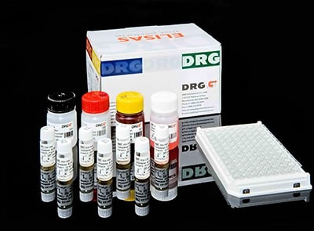
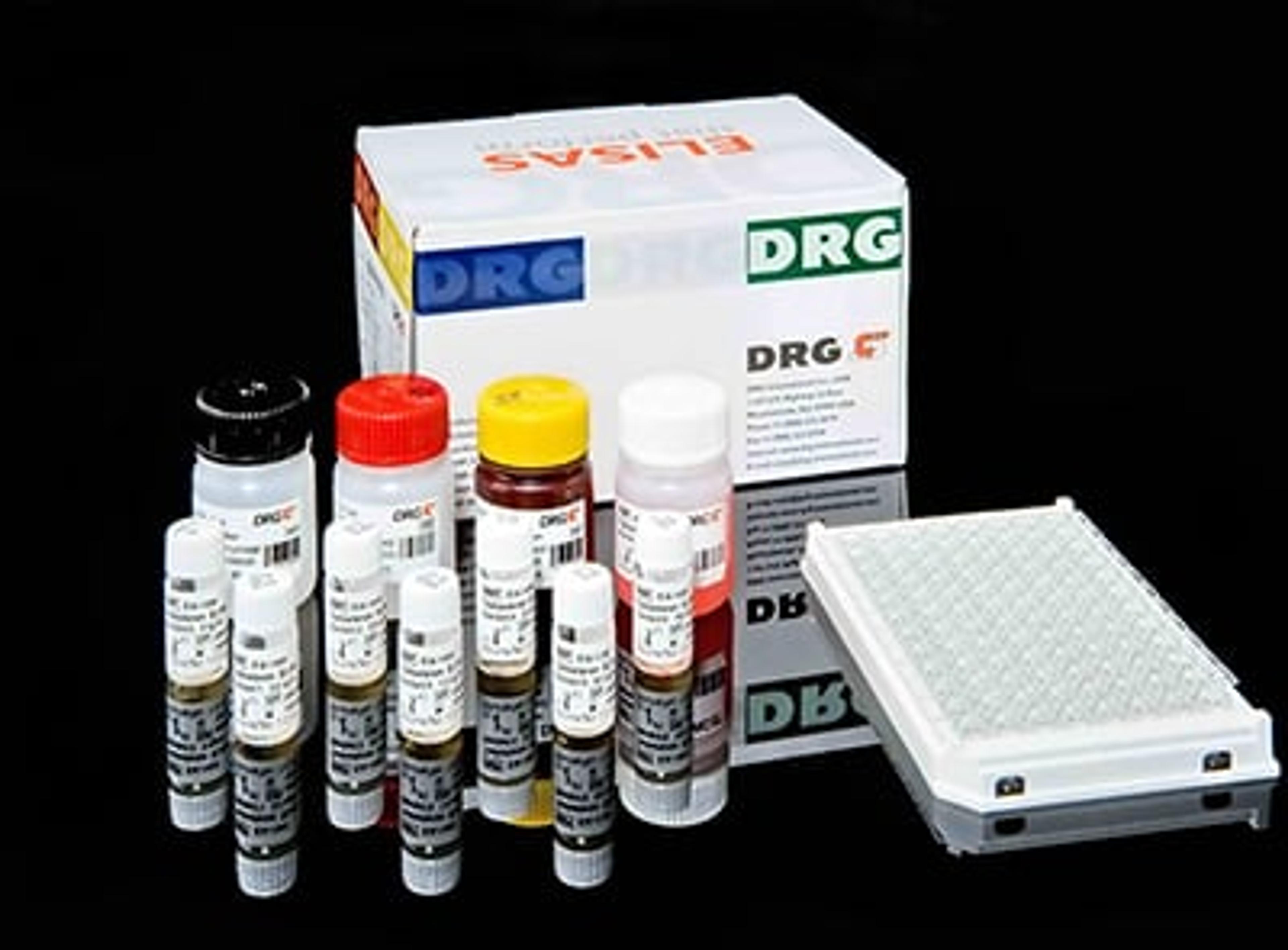
The VMA ELISA is designed for in vitro quantitative measurement of vanillylmandelic acid (VMA) concentration in patients’ urine.For in vitro diagnostic use only. Catecholamines include dopamine (found mostly in the central nervous system), norepinephrine (mainly in the sympathetic nervous system) and epinephrine (mainly in the adrenal medulla). They are stored as inactive complex. Released catecholamines, having a short half-life, are taken up by sympathetic nerve endings, or metabolized by the liver and kidney and excreted. Vanillylmandelic acid (VMA) and 4-hydroxy-3-methoxy-mandelic acid (HMMA) are the end product of both epinephrine and norepinephrine catabolism. Quantitation of the acidic metabolites has long proven to be a reliable diagnostic as well as commonly used follow-up procedure for pheochromocytoma and other catecholamine-,producing tumors (14-16). The prevalence of pheochromocytoma is 0.1% to 0.2% of hypertensive patients (17, 18). Pheochromocytomas have a long record of misdiagnosis due to the metabolic, cardia and gastrointestinal symptoms that can mimic many other diseases. Undetected, or mistreated it can be fatal. However, since this is a surgically curable disease, early diagnosis by demonstration of excess VMA excretion is critically important.The VMA ELISA is a solid phase enzyme-linked immunosorbent assay (ELISA) based on the competition between VMA coated on a microtiter well and that in urine for the monoclonal antibody. Outlined steps are:1. Sampling and reaction: The samples are incubated in the wells with horseradish peroxidase conjugated anti-VMA monoclonal antibody.2. Washing: Unbound VMA and the antibody bound to urinary VMA are removed by washing with 0.9% NaCI solution.3. Enzyme Reaction (Color Development): The amount of bound peroxidase is inversely proportional to the concentration of the VMA present in the urine sample. Upon addition of the substrate (TMB), a blue color is developed, then it is changed to yellow by adding Stopping Solution. The intensity of this is inversely proportional to the concentration of VMA in the Calibrator or urine sample.4. Absorbance Detection: After addition of Stopping Solution, absorbance is measured at 450 nm. And the readings are converted into the concentrations from the Calibration curve.

