Wire Myography
Wire myography is an in vitro technique that allows us to examine functional responses and vascular reactivity of isolated small resistance arteries. Vessels from various species, including transgenic models, and vascular beds can be examined in a variety of pathological disease states. Vessels are dissected, cleaned, and then mounted onto a channel myograph under isometric techniques. Each vessel is then normalized to determi…
Wire myography is an in vitro technique that allows us to examine functional responses and vascular reactivity of isolated small resistance arteries. Vessels from various species, including transgenic models, and vascular beds can be examined in a variety of pathological disease states. Vessels are dissected, cleaned, and then mounted onto a channel myograph under isometric techniques. Each vessel is then normalized to determine maximum active tension development. This allows the standardization of initial experimental conditions, an important consideration when examining pharmacological differences between vessels. Learn more about Living System’s wire myograph options below.
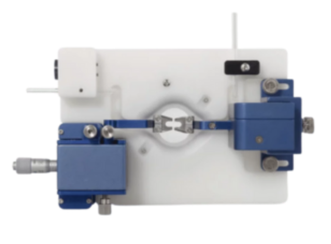
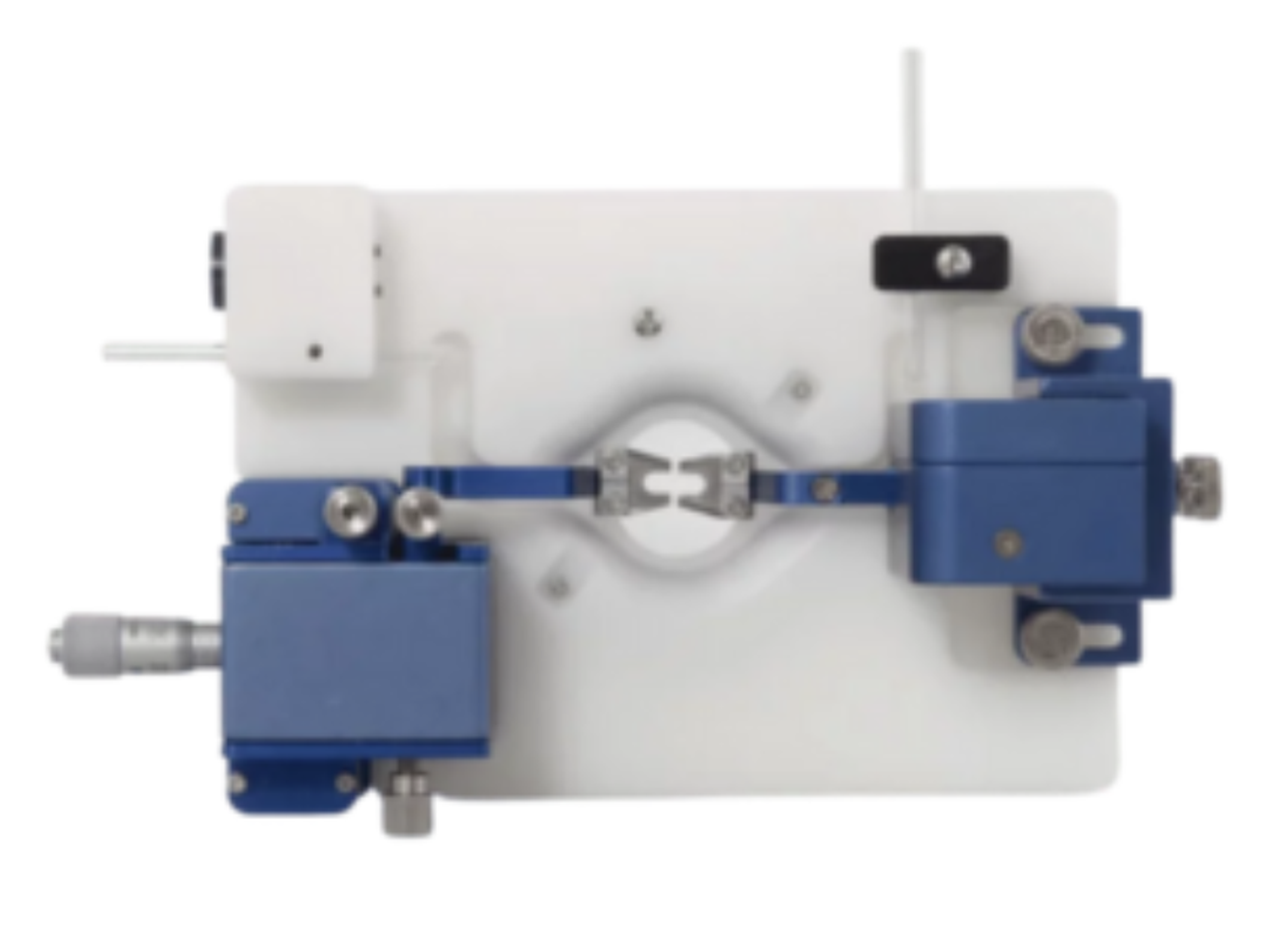
The classic Halpern/Mulvany style wire myograph is back as a standard product available from Living Systems Instrumentation. Living System’s MYO-CH wire myograph chamber is faithful to the basic design first introduced by Living Systems Instrumentation founder, the late Professor William Halpern, and Professor Michael Mulvany in their classic 1976 Nature paper titled “Mechanical properties of vascular smooth muscle cells in situ” (Nature 260(5552): 617-619, 1976). At the same time, state-of-the-art materials, machining, and force technology have been introduced.
Living System’s wire myographs include three types of tissue supports: wire-mounts for microvessels, L-bars for large ring preparations, and a hook for working with strips. Thus, Living System’s myograph is suitable for a range of applications including force measurements in microvessel rings, large vessels such as carotid artery and aorta, airway, intestine, bladder, and many more.
- Modular tissue supports that allow use of wire-mount microvessel ring preparations, Large and Small L-bars for working with large and small ring preps, and a hook attachment for working with strip preparations.
- Convenient linear position controls in all three axes for the fixed tissue support. Force transducer support features smooth up-down position control, which simplifies alignments and initial slack adjustments.
- Small bath volume (~5 ml) and optional self-heating capability for efficient use of expensive peptides and reagents.
- Convenient integrated flow path helps achieve optimal flow pattern when bath superfusion is used.

