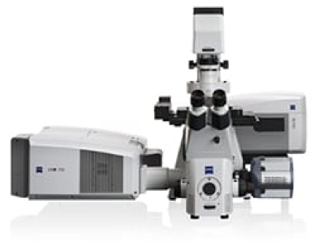
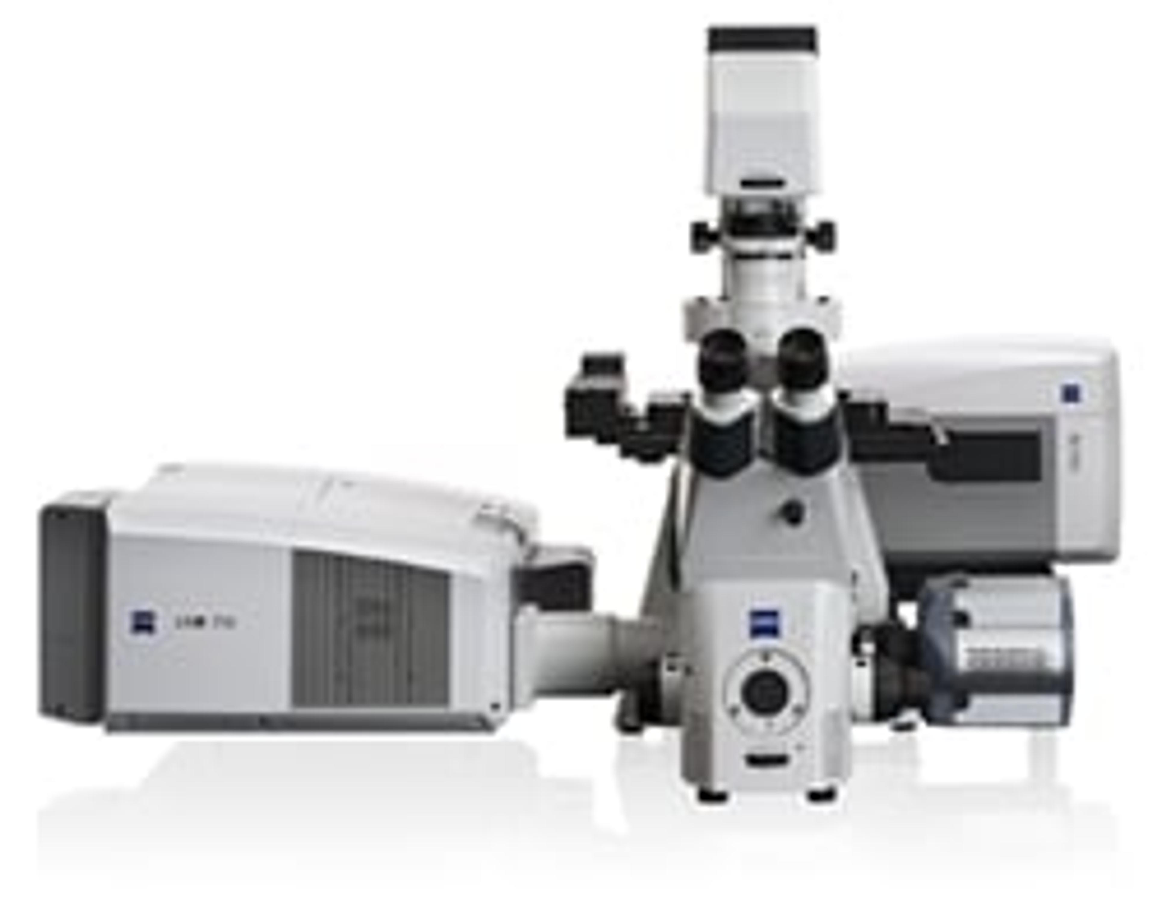
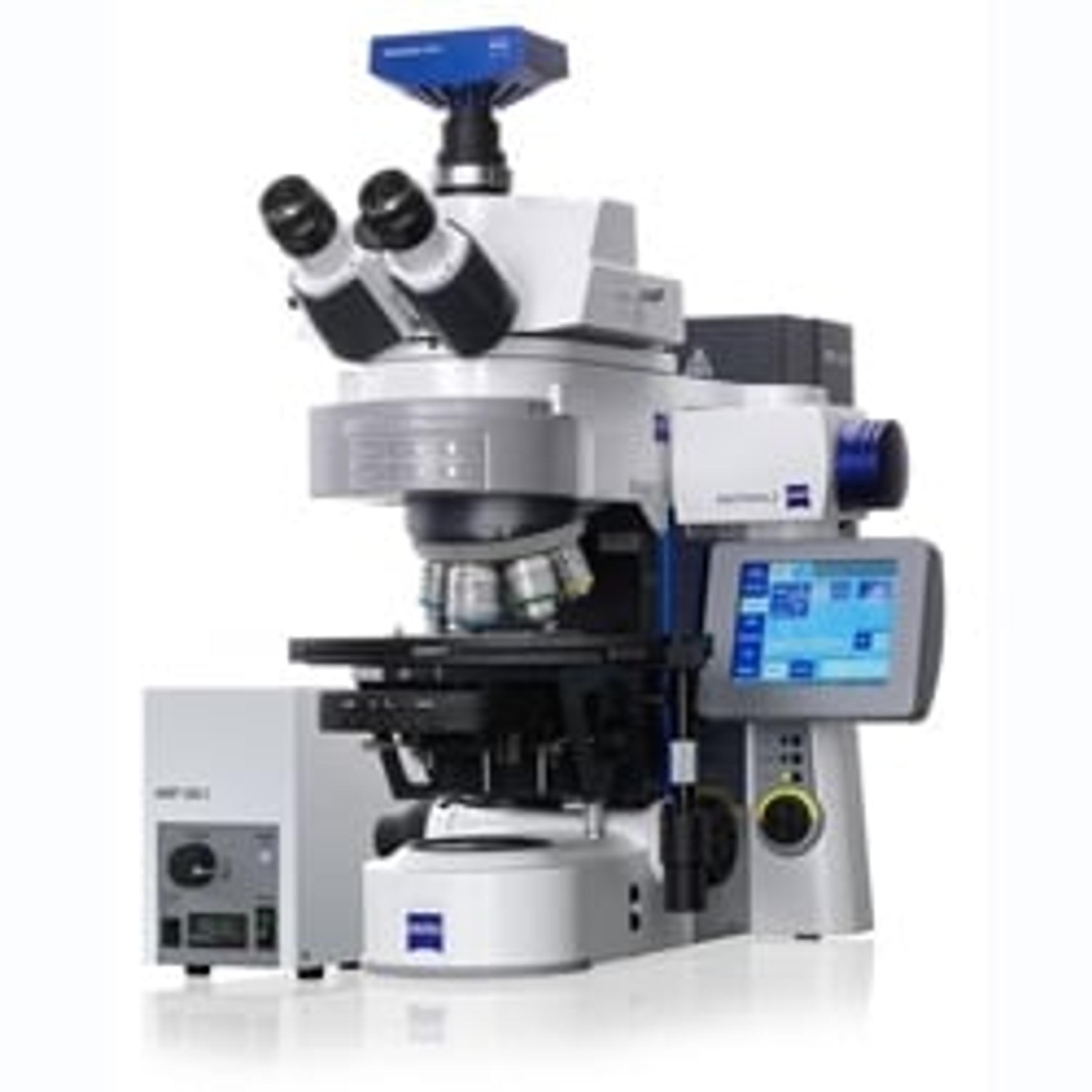
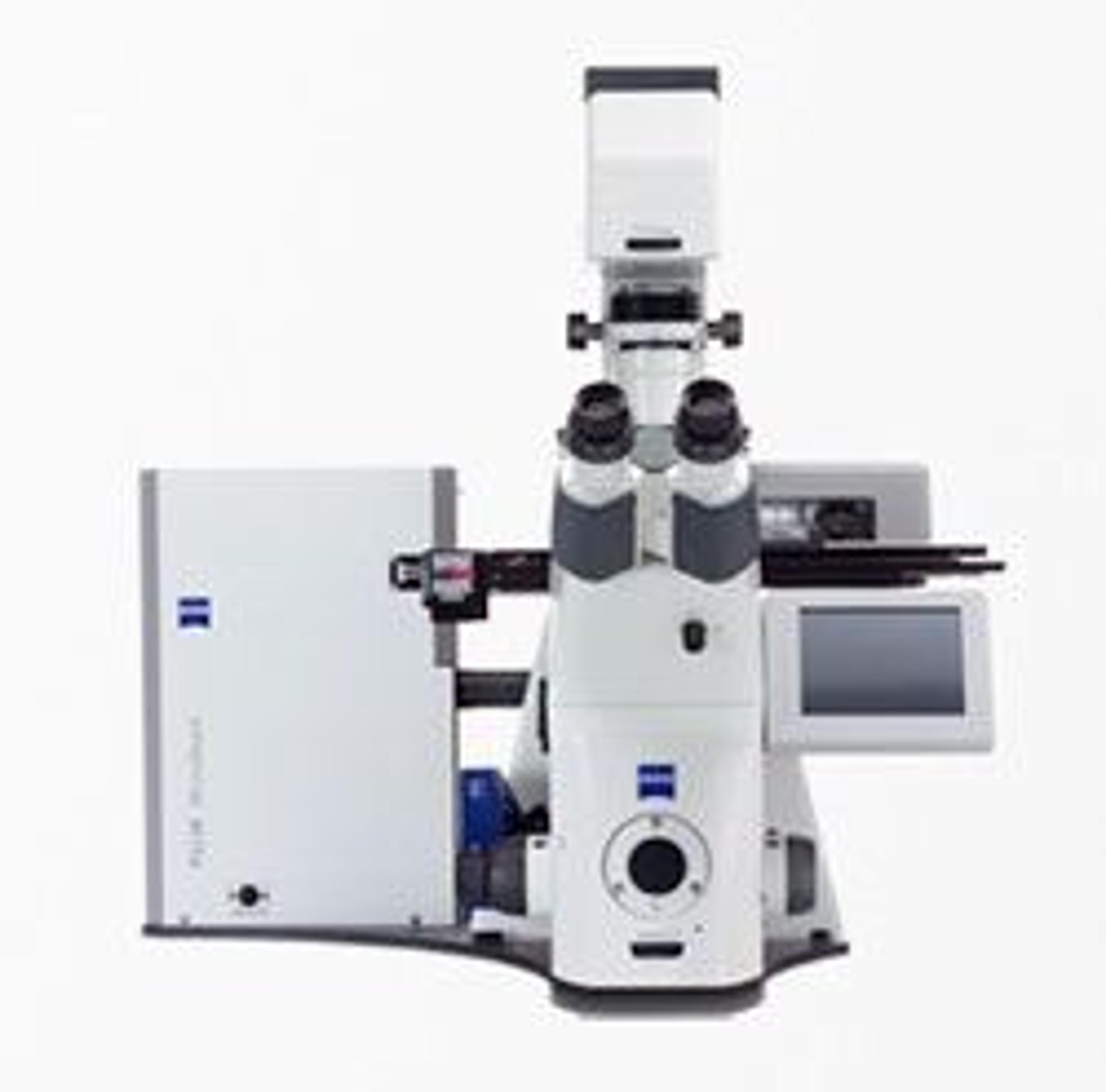
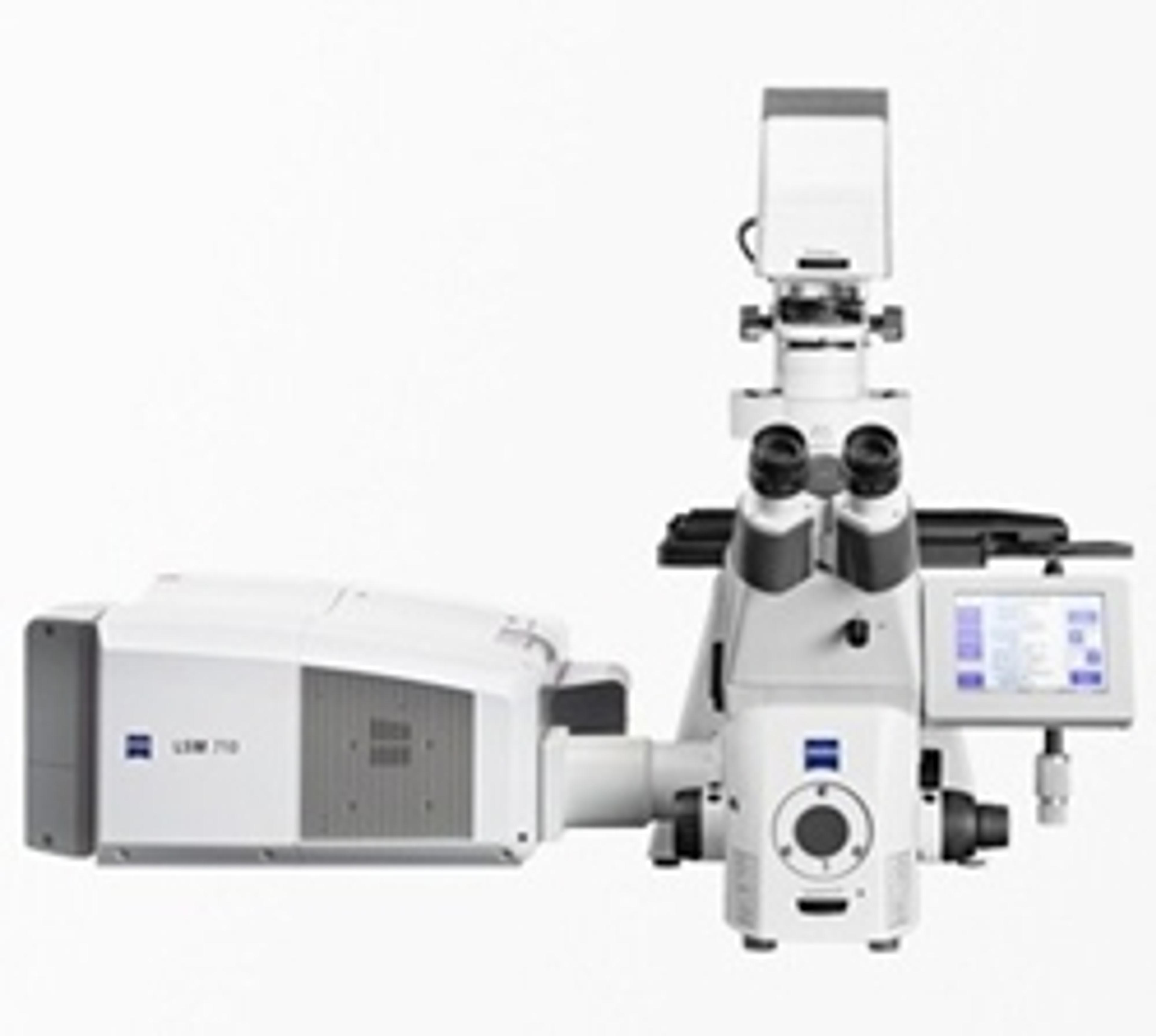
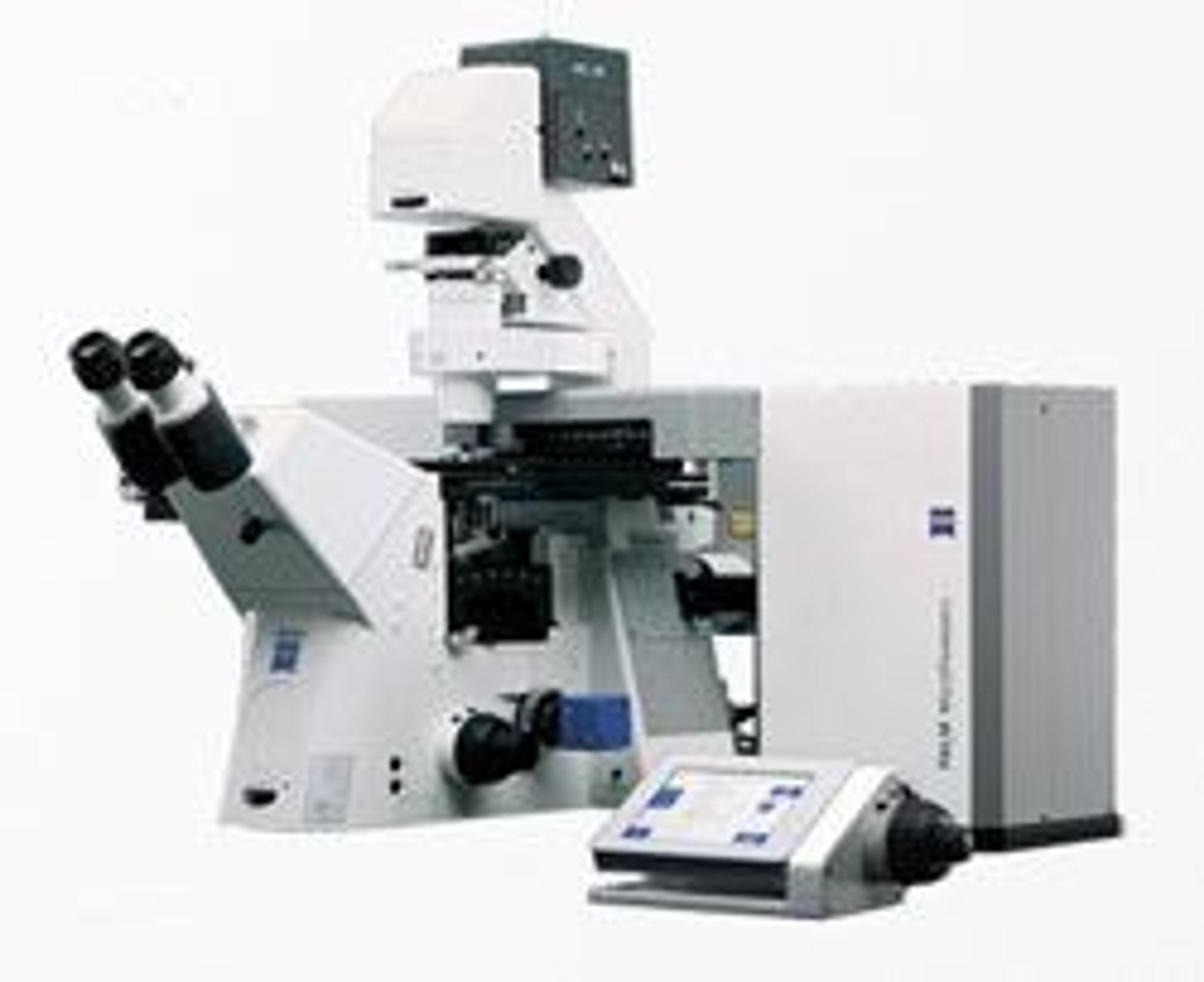
ZEISS ELYRA S.1
Put Flexibility First with Structured Illumination. ELYRA S.1 images any fluorophore – with up to twice the resolution of a conventional light microscope. Using structured illumination (SR-SIM) you reveal fine structural details while remaining free to label your samples with conventional dyes.You have invested a lot of time and energy in producing fusion proteins and multicolor staining protocols that are adapted to your exp…
Put Flexibility First with Structured Illumination.
ELYRA S.1 images any fluorophore – with up to twice the resolution of a conventional light microscope. Using structured illumination (SR-SIM) you reveal fine structural details while remaining free to label your samples with conventional dyes.
You have invested a lot of time and energy in producing fusion proteins and multicolor staining protocols that are adapted to your experimental system. Now, with ELYRA S.1, you capture superresolution microscopy data with ease, using samples that may already be available in your lab's freezer. Specially-designed gratings give you the best resolution for each wavelength. Do you need Z-sectioning for 3D data acquisition? A fast, light-efficient detection? Then ELYRA S.1 is your ideal choice.
Applications:
- Resolve structural detail in 3D with high penetration depth.
- Probe the structural organization of a whole cell.
- Investigate arrangement of cellular components and proteins.
- Explore interaction of molecules.
- Reveal the ultrastructure of organelles.
- Probe the ultrastructure of molecular assemblies.
- Map protein localization onto a structural context.
- Track many molecules and retrieve diffusion behavior.
- Study structural changes of slower dynamics.

