Western Blotting Products & Reviews
25
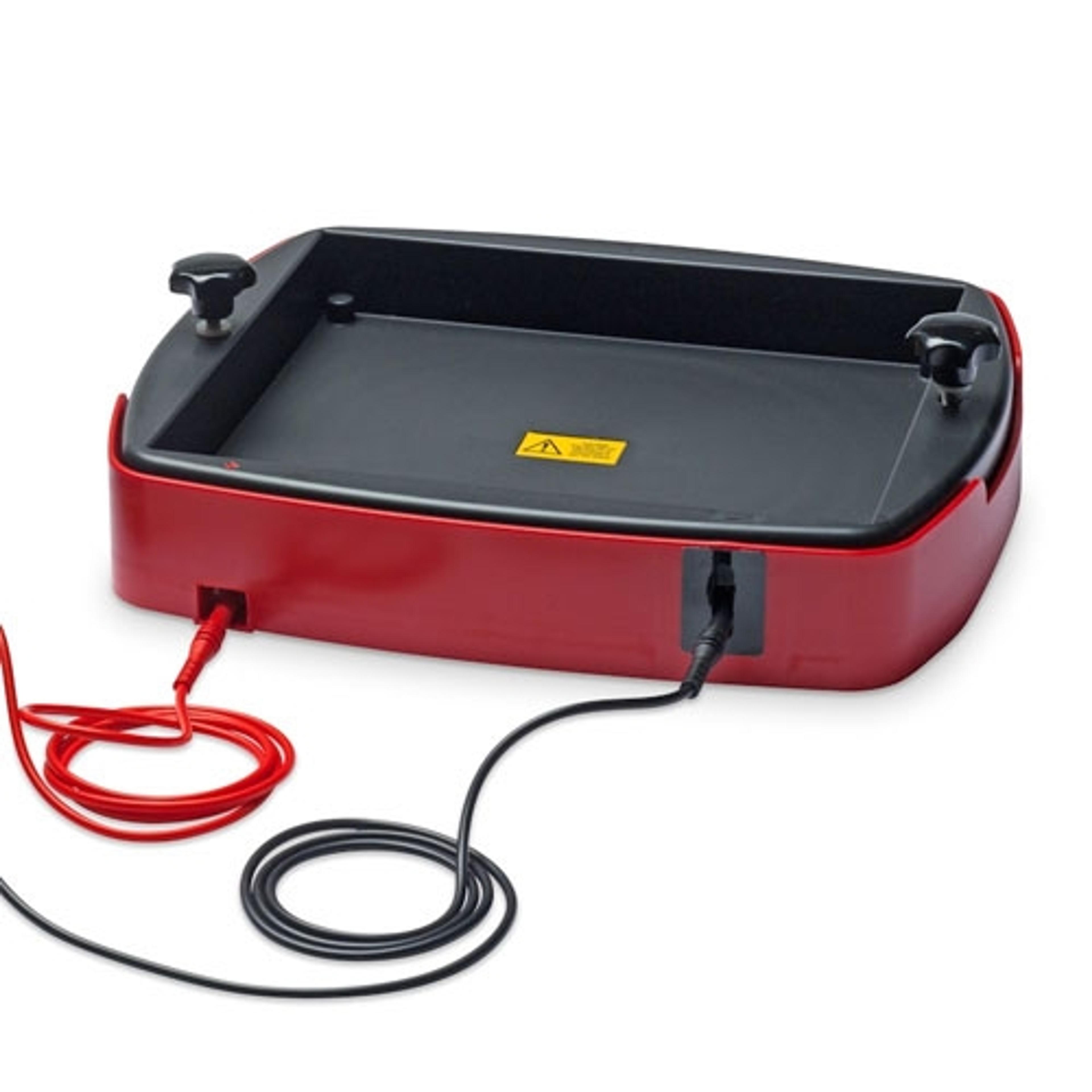
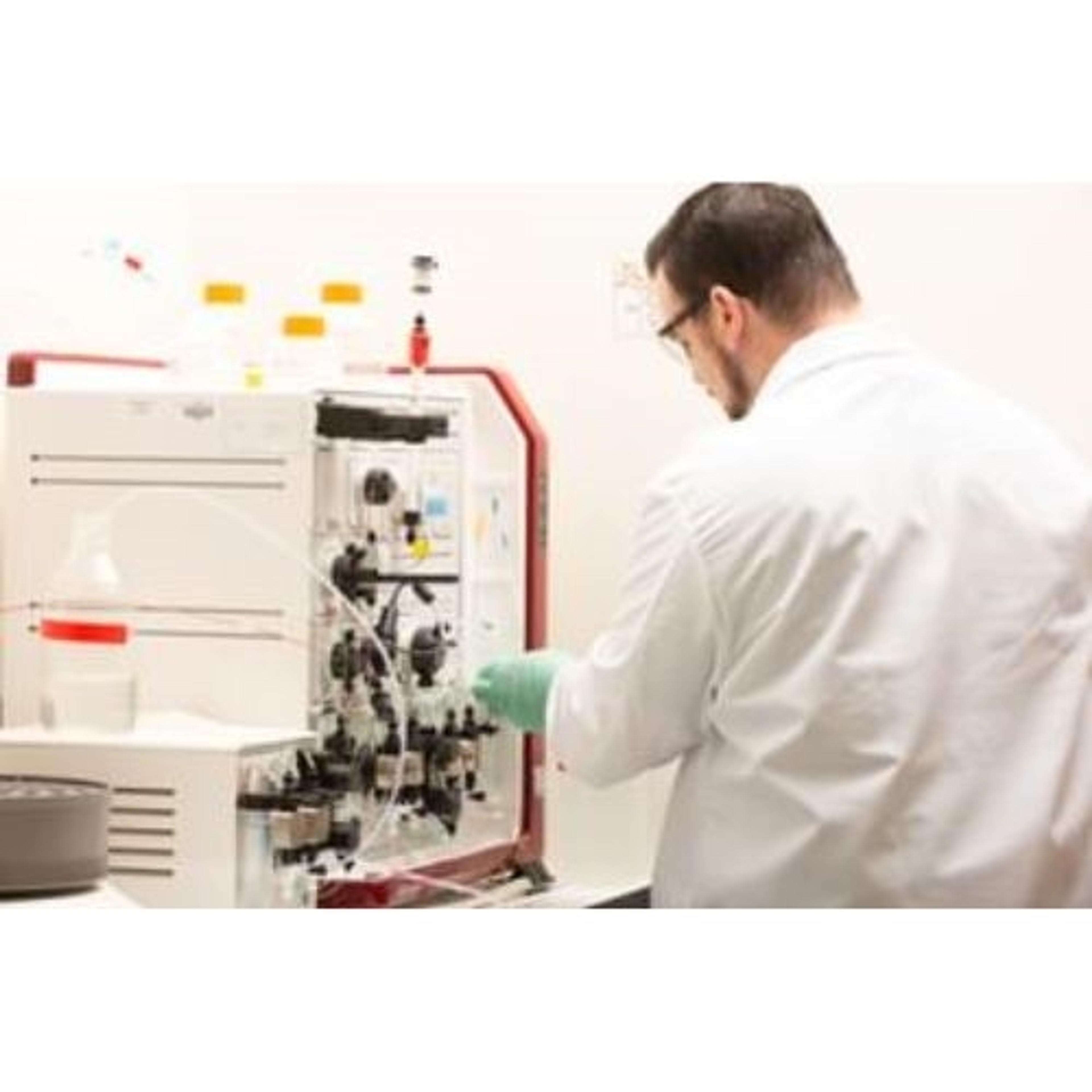
Western blotting equipment is used to transfer and identify specific proteins within a sample, reveal protein modifications, as well as give a semi-quantitative estimation of their concentration. Western blotting equipment includes all apparatus necessary to transfer proteins from gel to membrane and subsequent processing steps. Protein transfer can be performed by electroblotting with wet, semi-dry and dry transfer systems onto nitrocellulose and PVDF membranes. Blocking, washing and labeling of membranes follows, involving buffers, blocking reagents, blotting / incubation trays, labeling reagents, immunoblotting assays, antibodies and conjugates. Automated equipment for these steps is available to accelerate your lab workflow. Finally, detection and imaging of proteins can be conducted using gel documentation and imaging systems. Find the best western blotting equipment in our peer-reviewed product directory: compare products, check customer reviews and receive pricing direct from manufacturers.
Selected Filters:
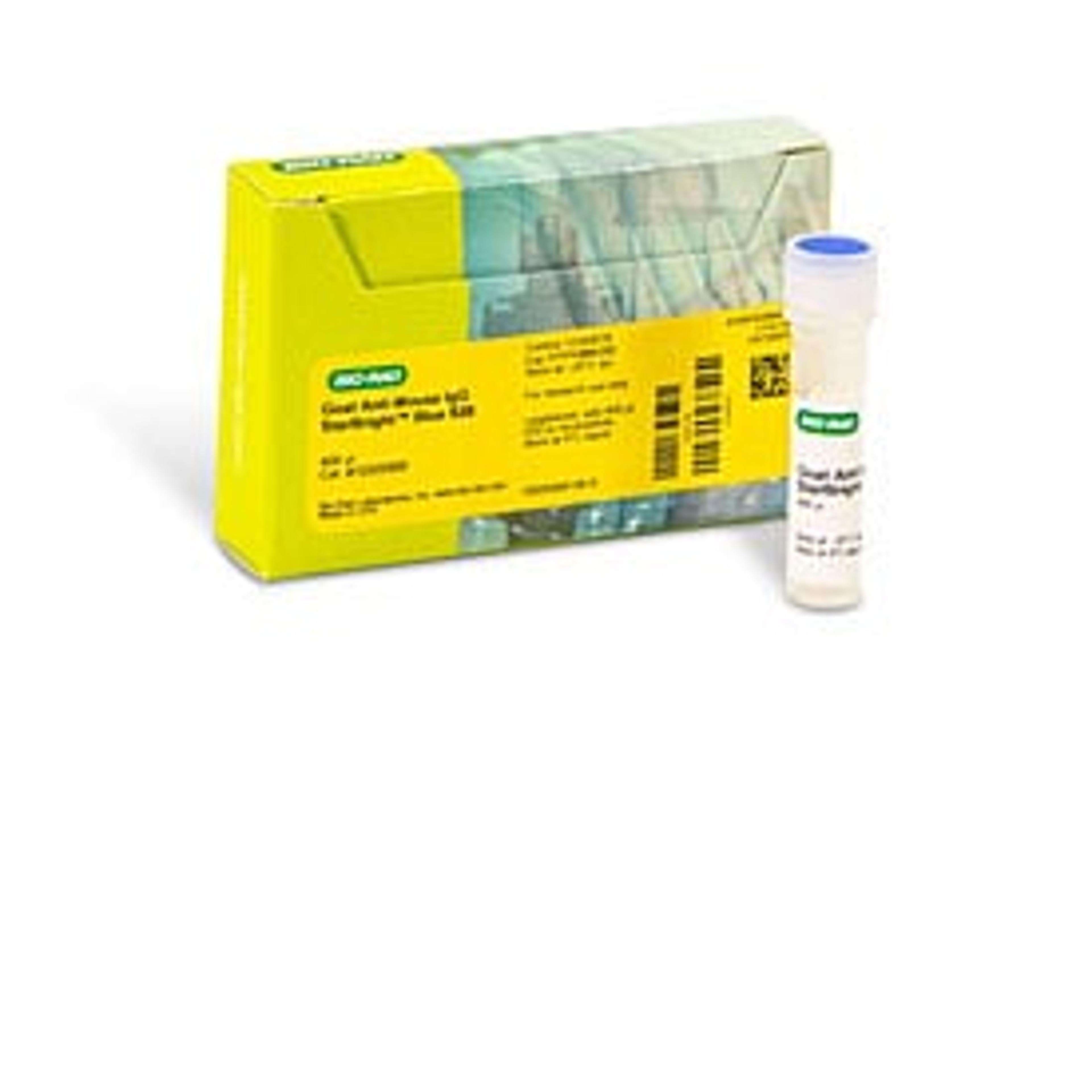
StarBright™ Blue 520 Goat Anti-Mouse IgG, 400 µl
Bio-RadPkg of 1, lyophilized goat anti-mouse IgG conjugated with StarBright Blue 520 Fluorophore; reconstitute to 400 µl

StarBright™ Blue 520 Goat Anti-Mouse IgG, 80 µl
Bio-RadPkg of 1, lyophilized goat anti-mouse IgG conjugated with StarBright Blue 520 Fluorophore; reconstitute to 80 µl
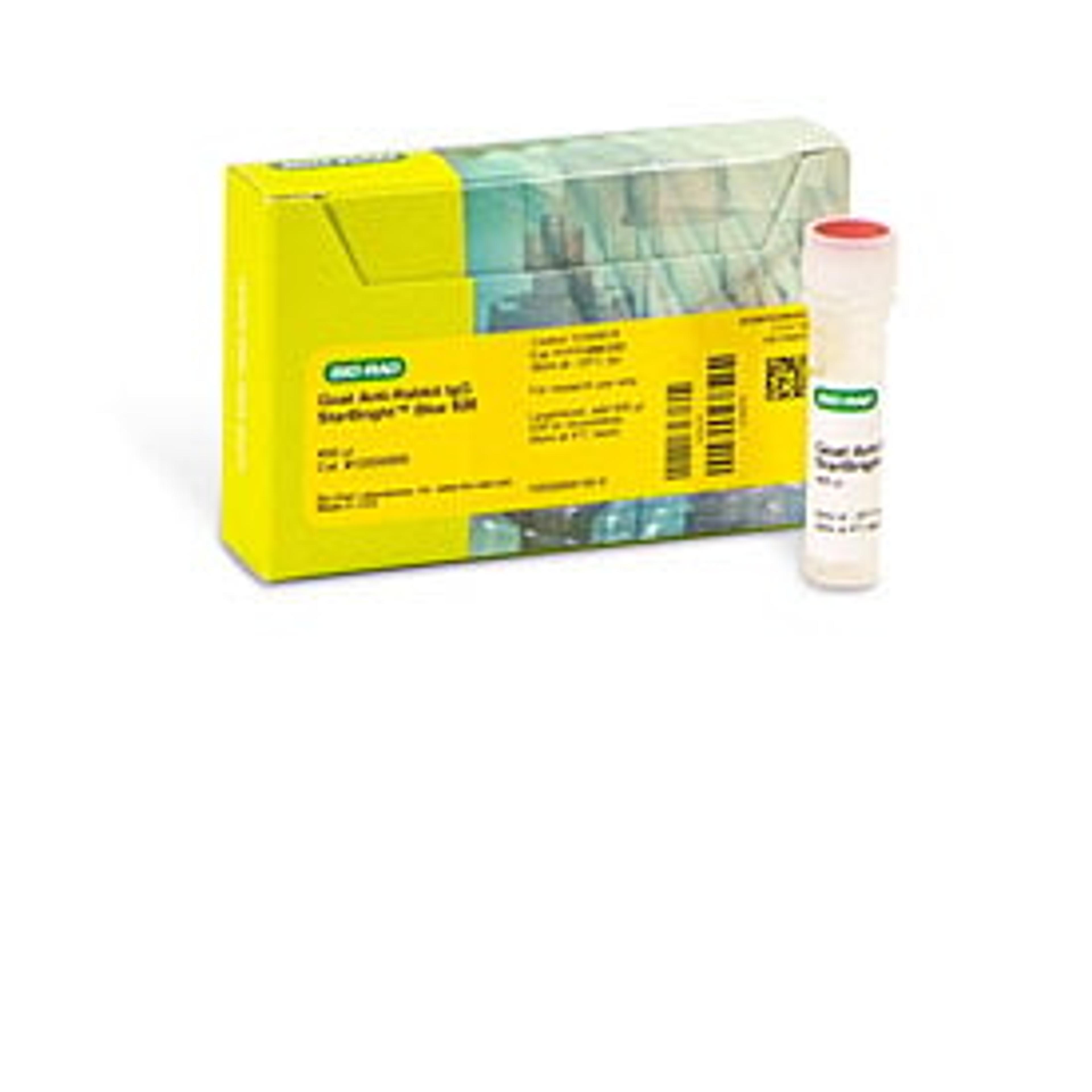
StarBright™ Blue 520 Goat Anti-Rabbit IgG, 400 µl
Bio-RadPkg of 1, lyophilized goat anti-rabbit IgG conjugated with StarBright Blue 520 Fluorophore; reconstitute to 400 µl
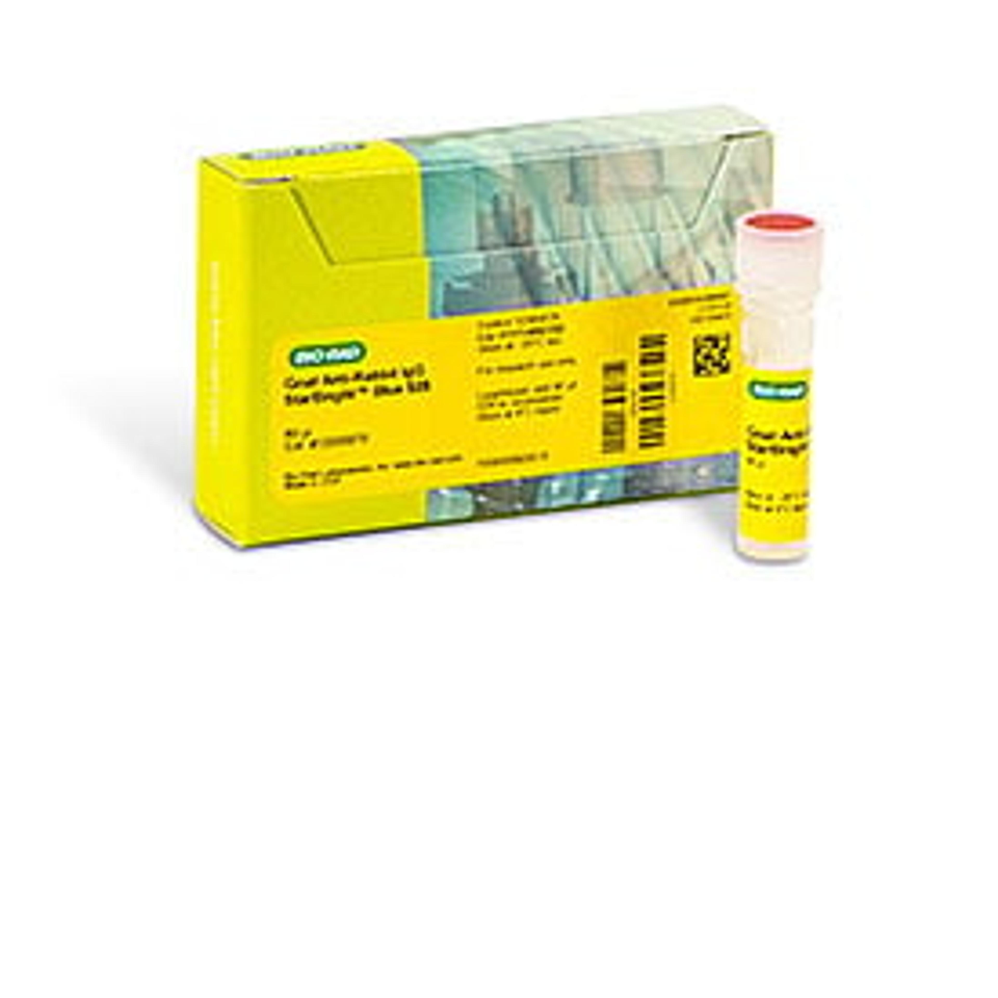
StarBright™ Blue 520 Goat Anti-Rabbit IgG, 80 µl
Bio-RadPkg of 1, lyophilized goat anti-rabbit IgG conjugated with StarBright Blue 520 Fluorophore; reconstitute to 80 µl
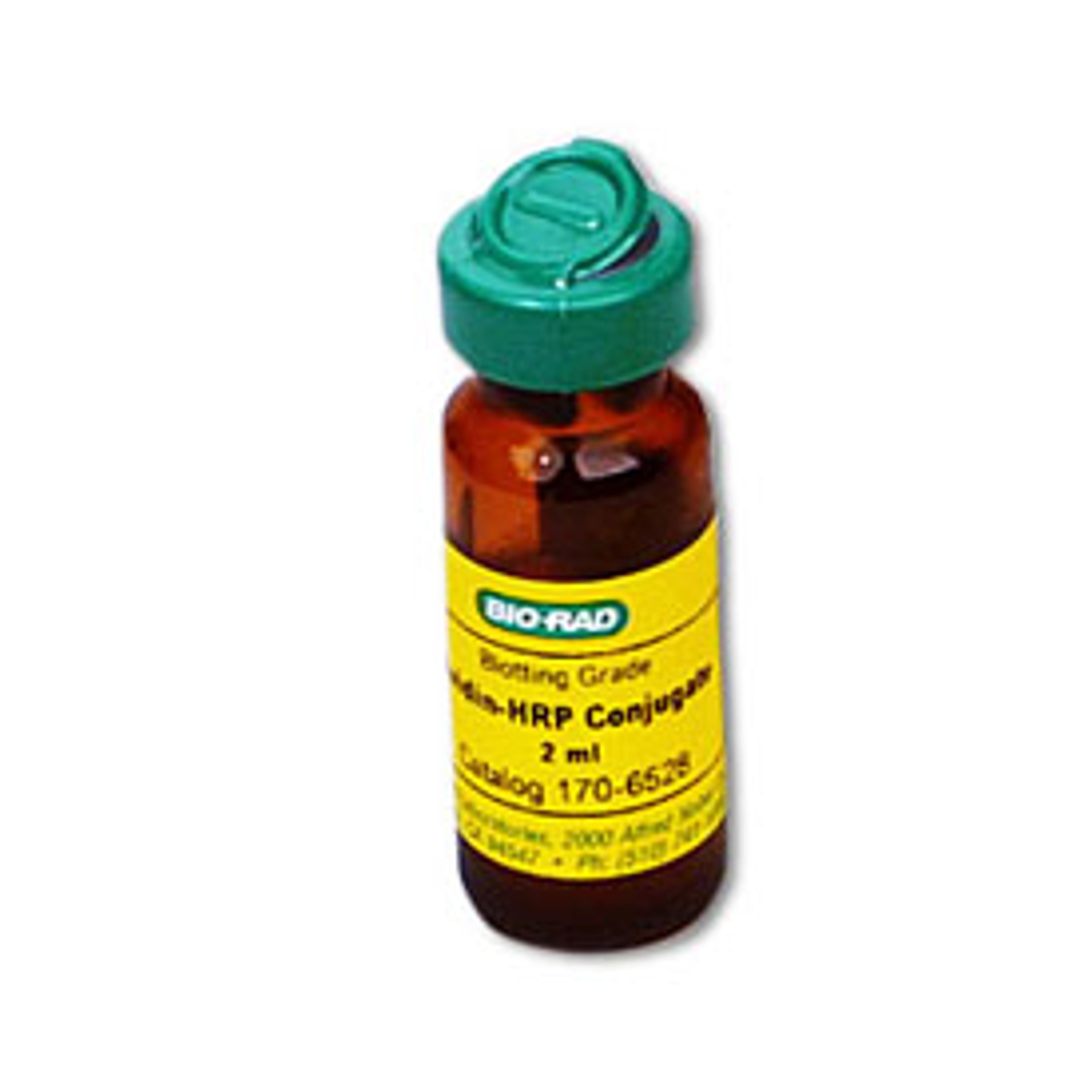
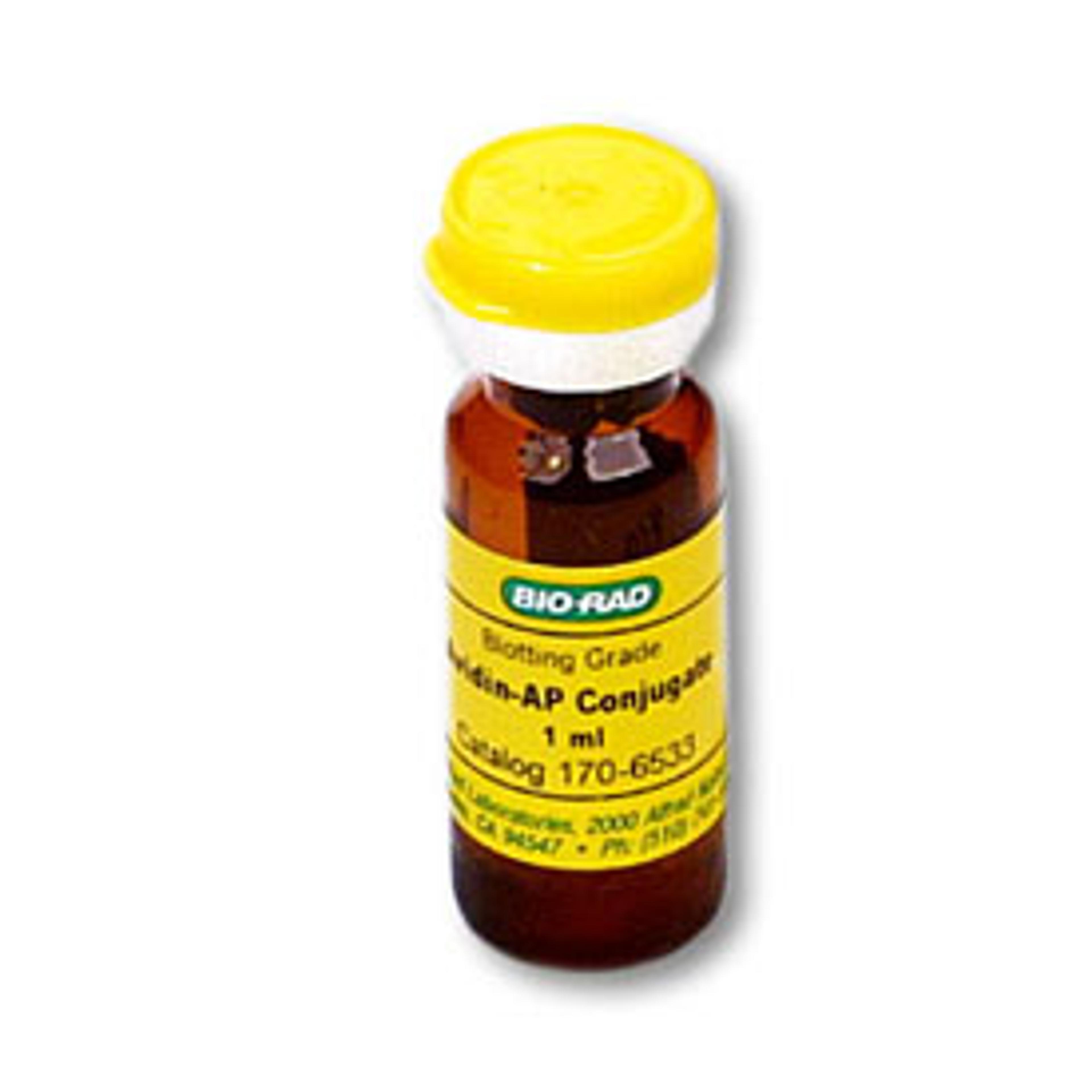
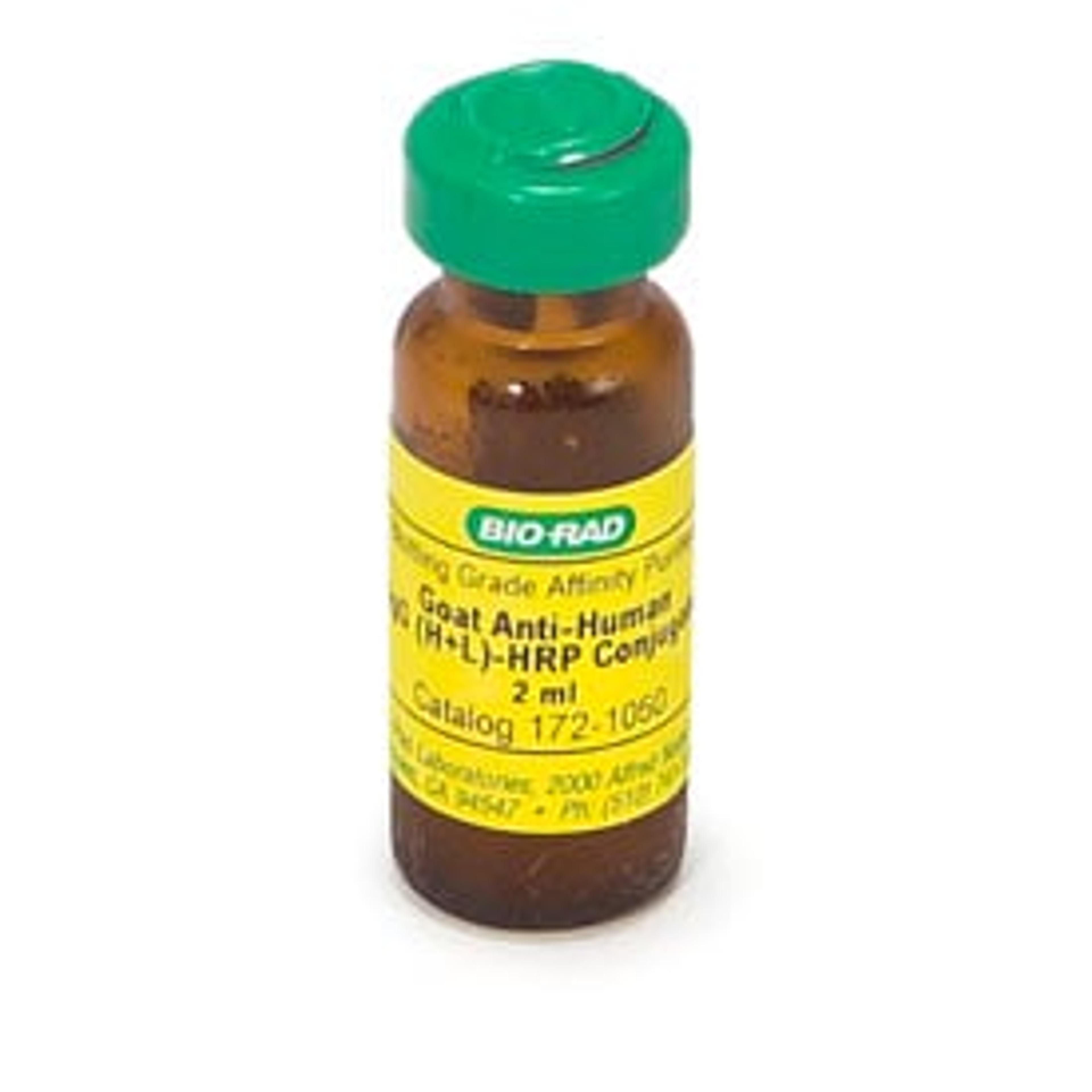
HRP and AP Conjugates
Bio-RadThe high titer of the blotting-grade antibody conjugates increase assay sensitivity. Greater working dilutions (1:3,000) decrease background and increase the signal-to-noise ratio of the assay.
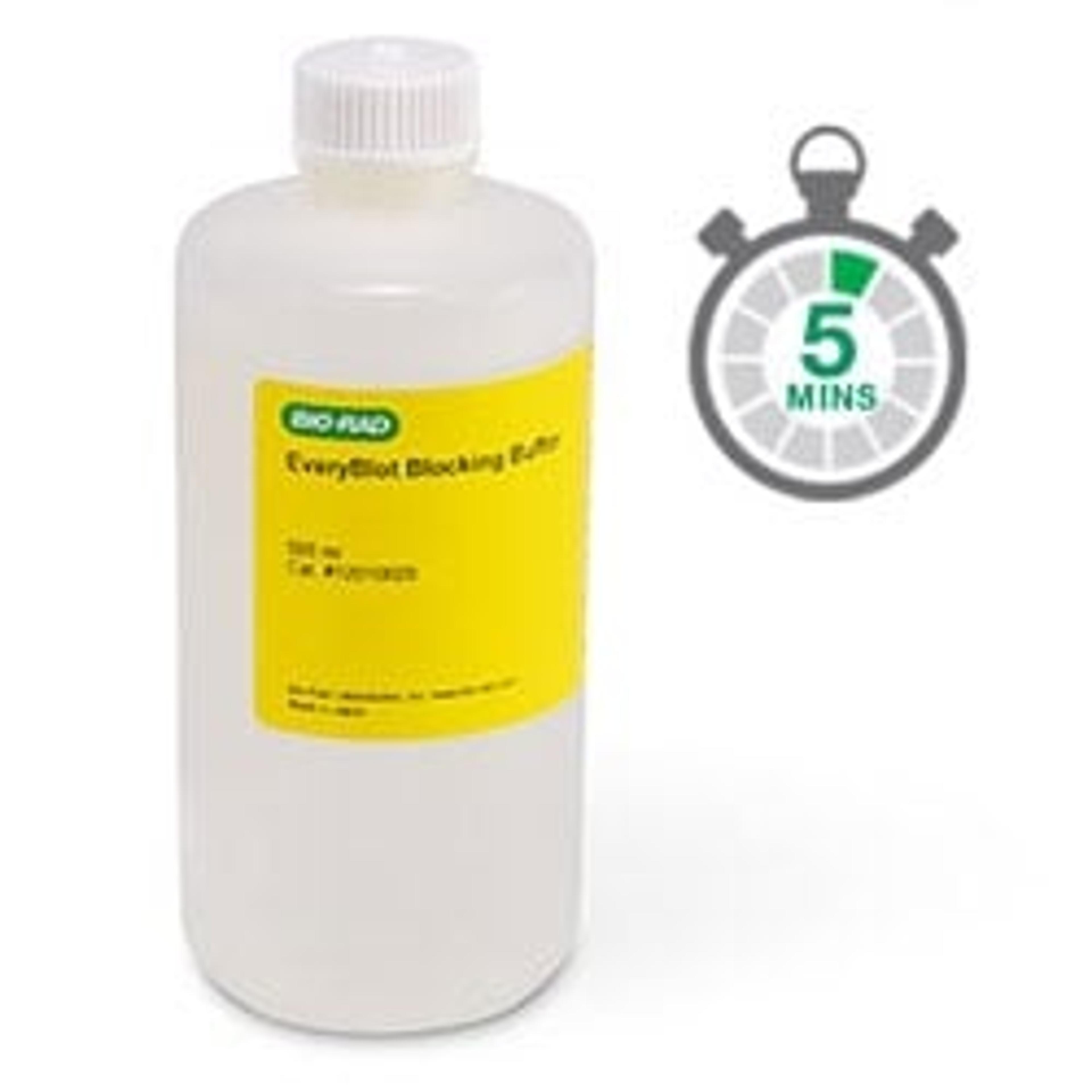
EveryBlot Blocking Buffer
Bio-RadEveryBlot Blocking Buffer provides 5 minute blocking and maximum sensitivity for all western blots regardless of detection method.
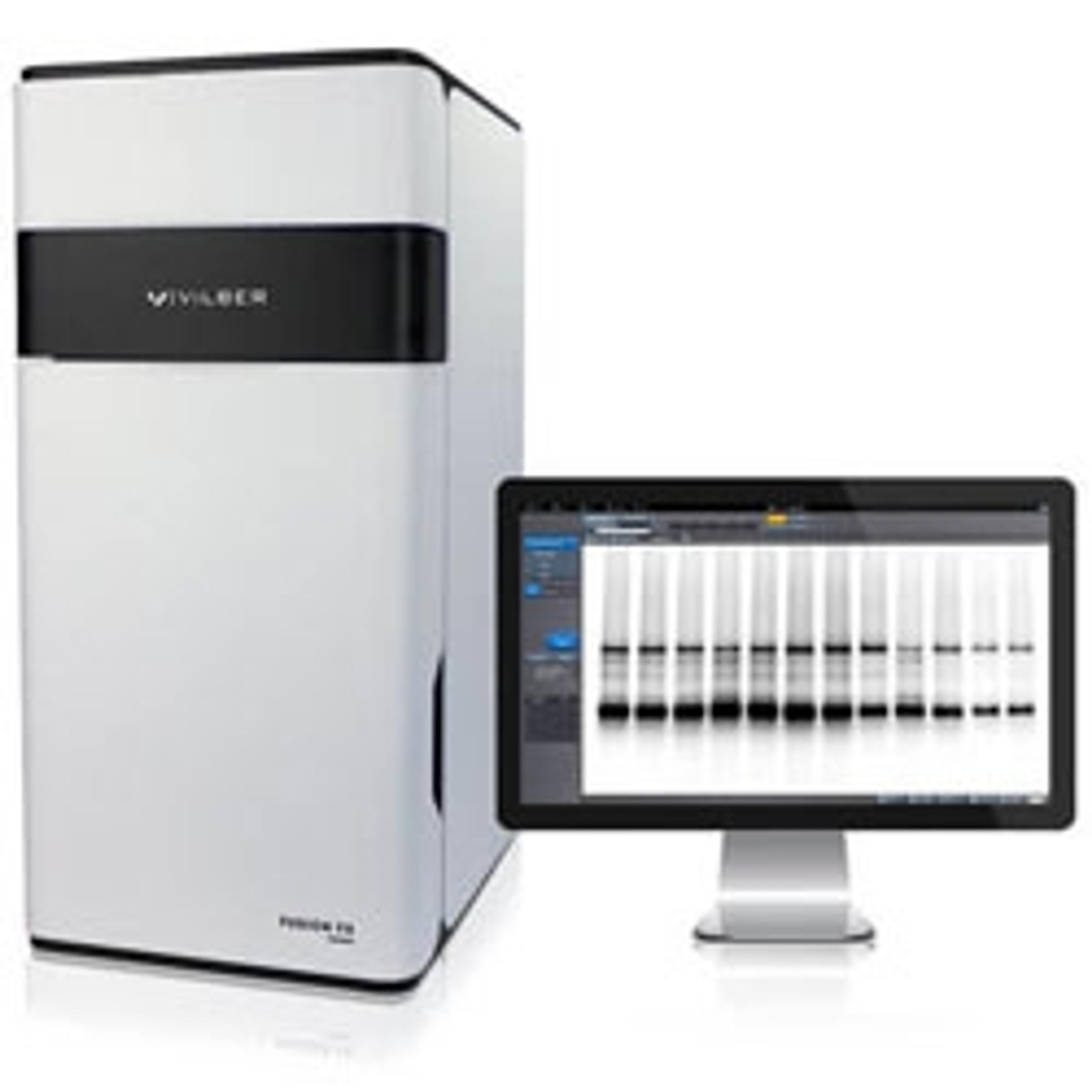
Vilber Fusion
Labtech InternationalFounded in 1954 Vilber are leaders in molecular imaging, having equipped more than 20,000 laboratories worldwide. Vilber Fusion blot imaging systems have been used by 3 Nobel prize winners. The latest generation of Fusion systems offer ‘better than film’ sensitivity with user upgradability for multi-colour fluorescence blotting.
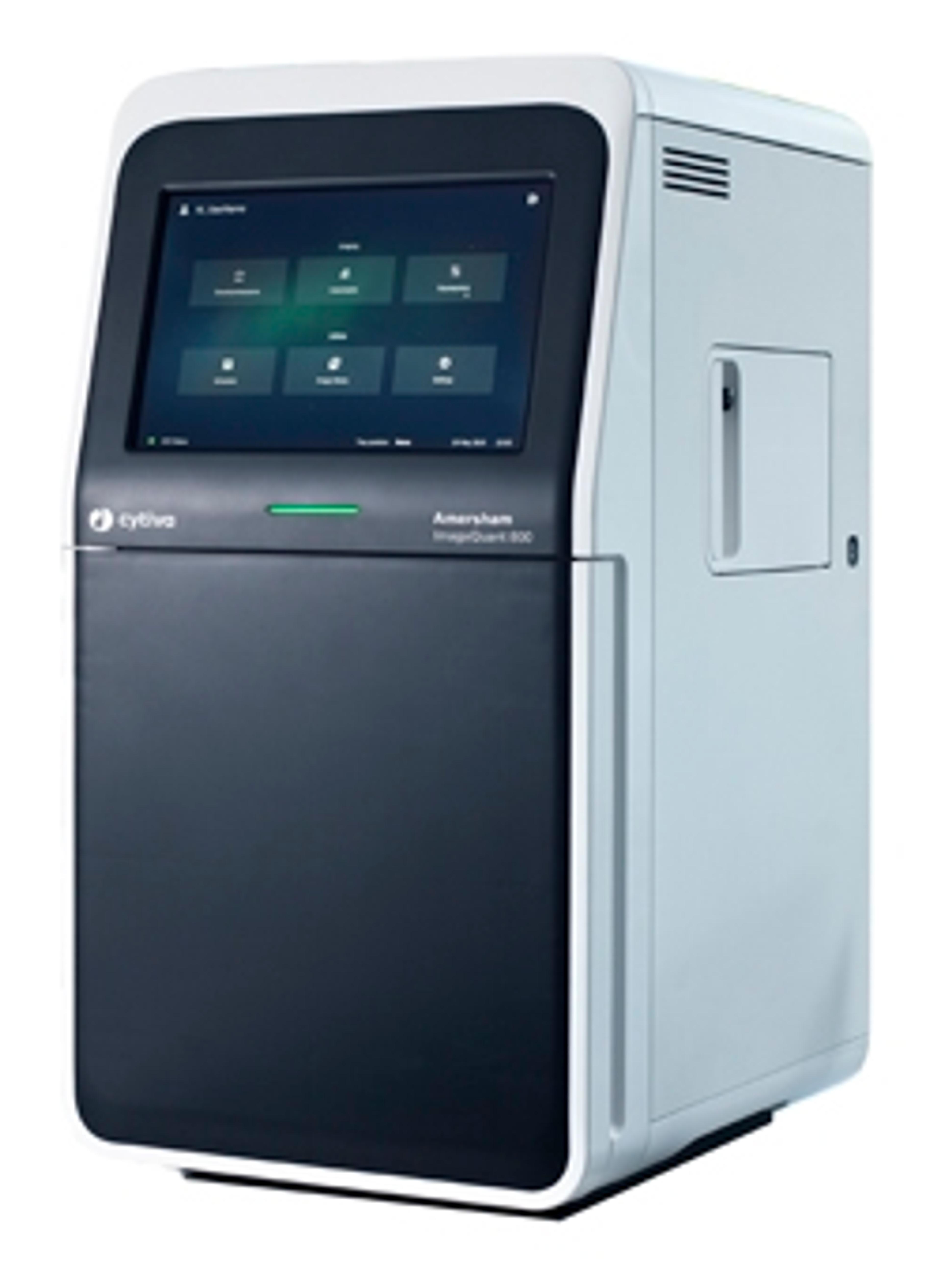
Amersham ImageQuant 800 systems
CytivaFlexible imagers with automated features for sensitive, accurate chemiluminescence and fluorescence detection. To support your electronic data management, an optional GxP module is available, designed to meet increasing requirements of regulations like FDA 21 CFR part 11 and EU GMP Annex 11.
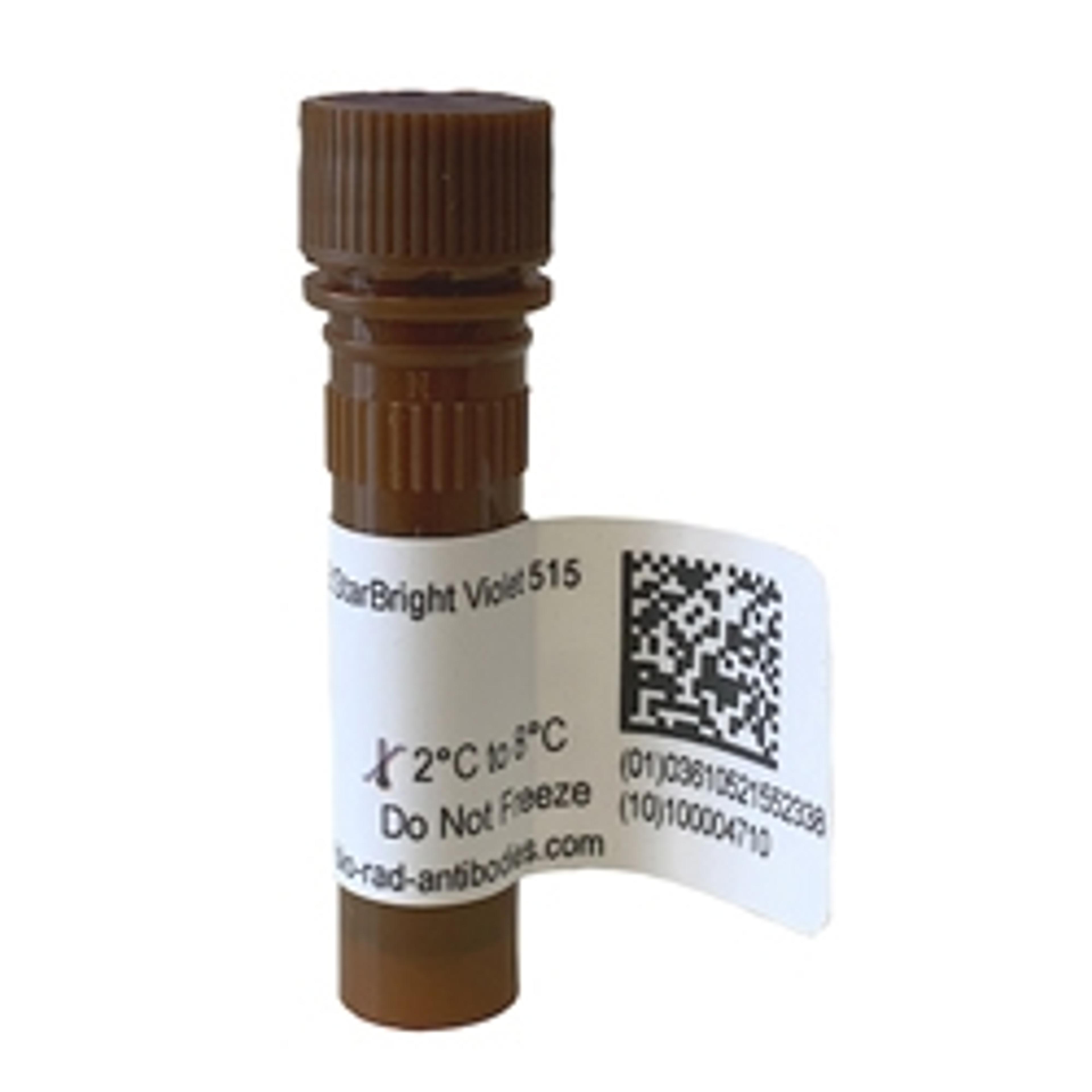
StarBright Violet 515 (SBV515) Dye
Bio-RadExcitable by the violet 405 nm laser and emitting at 515 nm, StarBright Violet 515 (SBV515) Dye is a perfect replacement for fluorophores that have emission maxima between 500 and 550 nm, such as Pacific Orange, Krome Orange, vioGreen, and Brilliant Violet 510, which can all be detected with a 525/50 filter (or similar) in your flow cytometry experiments. Minimal excitation by other lasers and a narrow emission profile make St…

Gold-in-a-Box™ Colloidal-Gold Conjugation Kits
AMSBIOFor preparing highly reactive antibody (purified) and protein (purified and soluble) gold conjugates
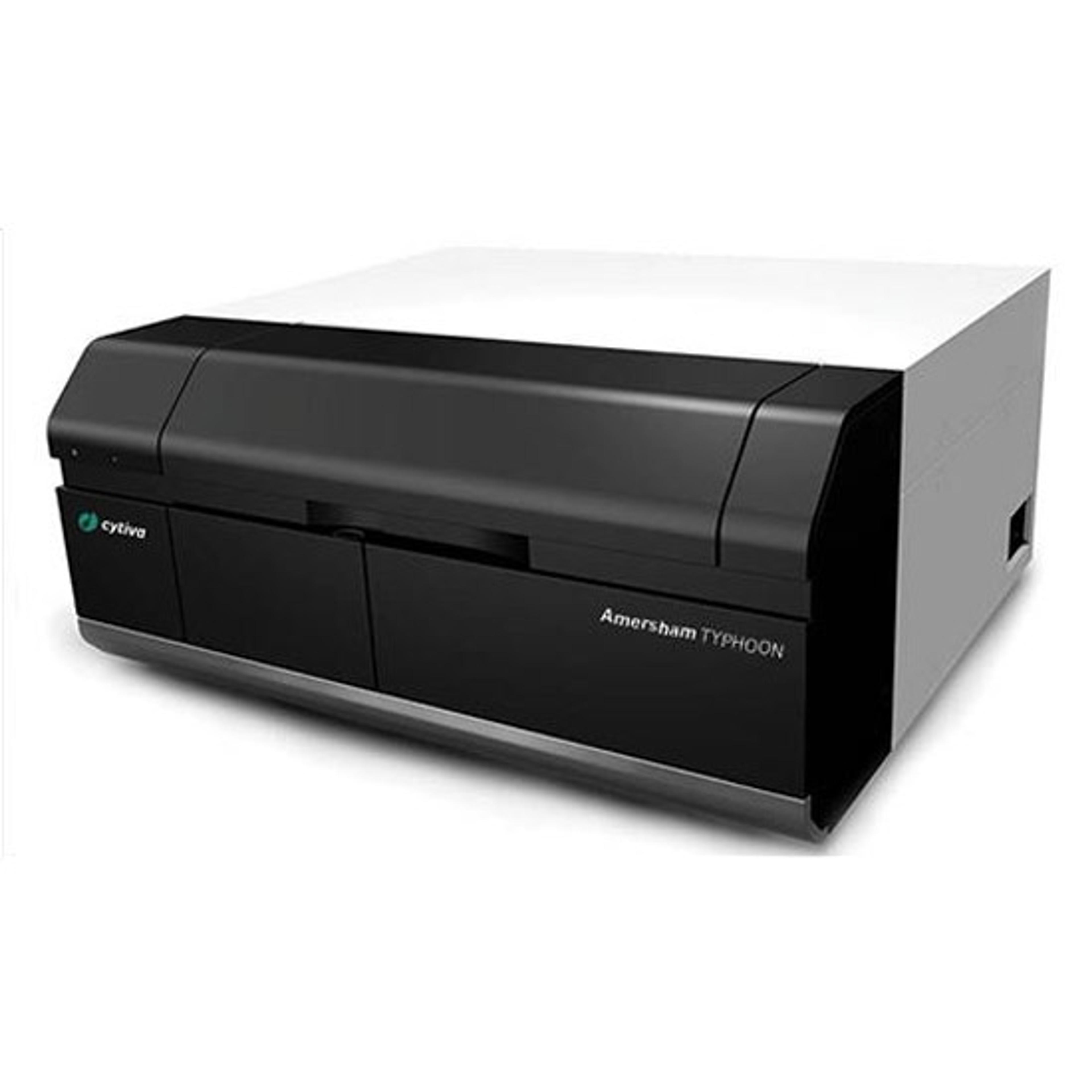
Typhoon laser-scanner platform
CytivaAmersham™ brand Typhoon™ laser-scanner platform provides exceptional data quality through extremely sensitive detection, high image resolution, and a very broad linear dynamic range. These versatile imaging systems support multiple imaging modes, including phosphor imaging, red/green/blue (RGB) and long and short wavelengths of near infrared fluorescence (NIR), as well as optical densitometry (OD) of proteins in stained gels.
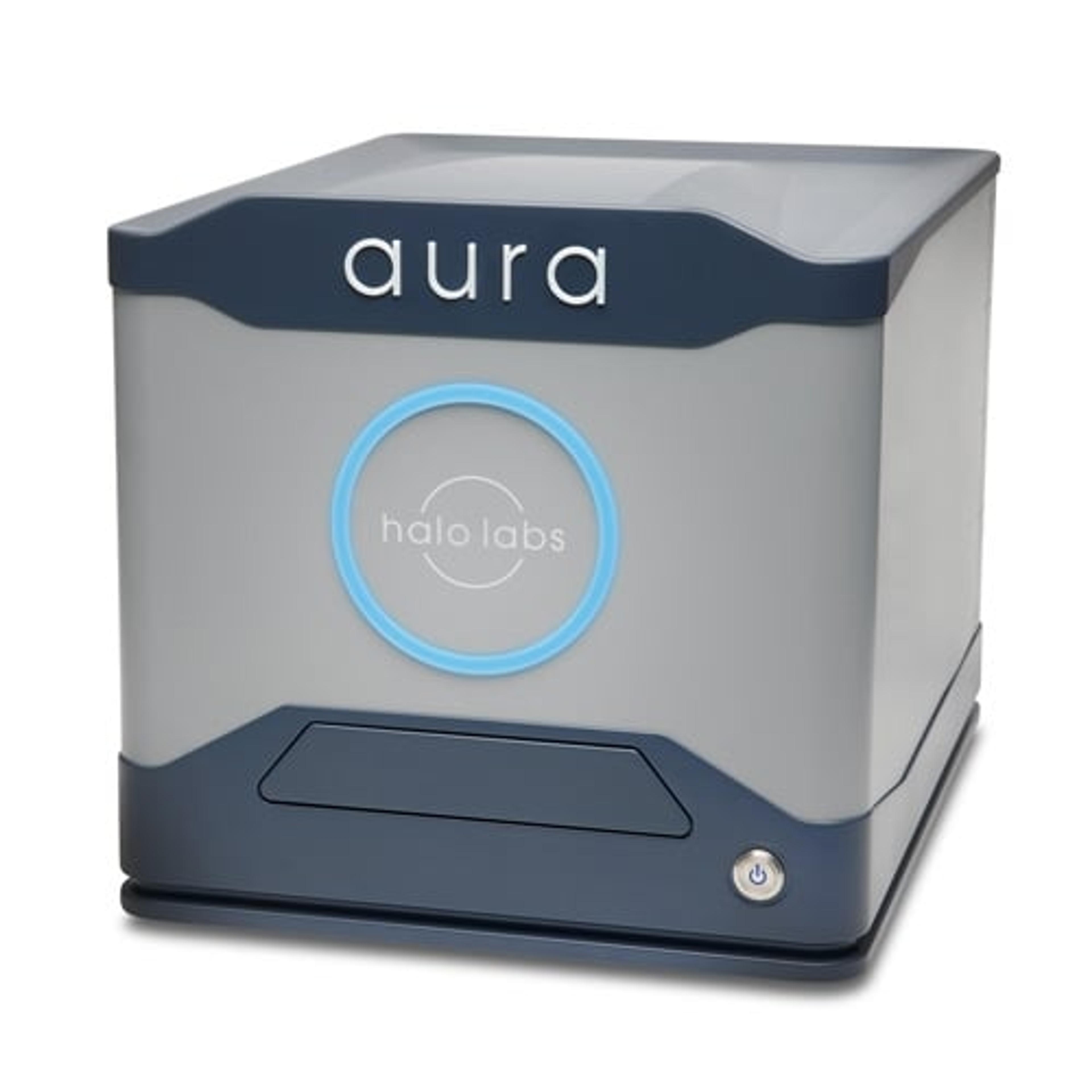
Aura
Halo LabsAura® is the particle and aggregate detection system that combines Backgrounded Membrane Imaging which images 100% of your sample to give you count, size, and morphological information, with up to 2 channels of Fluorescence Membrane Microscopy. Identify if particles are protein or not with the first FMM channel and determine if your sample contains lipids, hydrophobic entities, or other aggregates of your choice with a second…
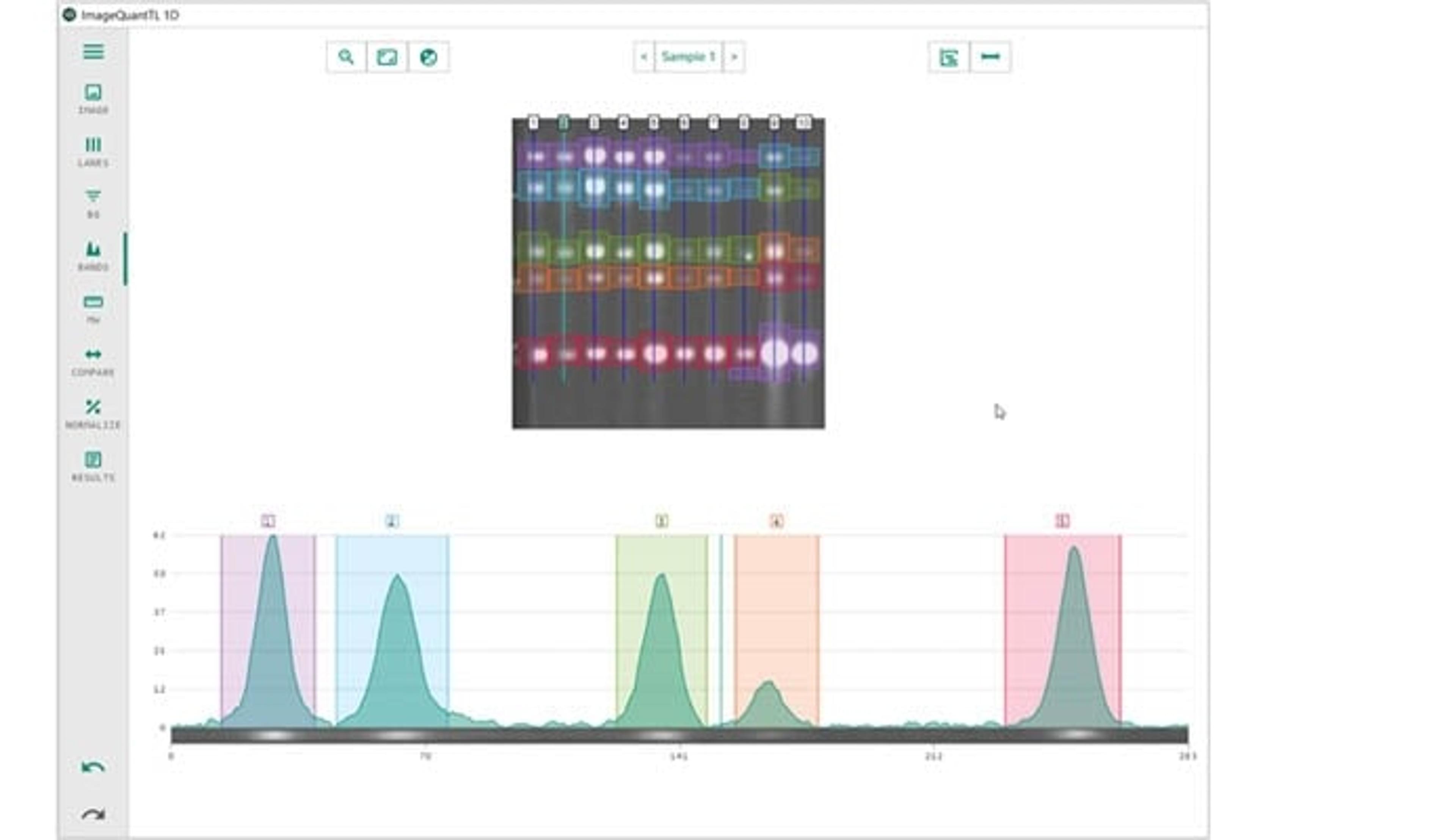
Amersham brand ImageQuant TL image analysis software
CytivaImageQuant™ TL is an advanced analysis software designed specifically for blots and 1D gel image analysis and quantification.
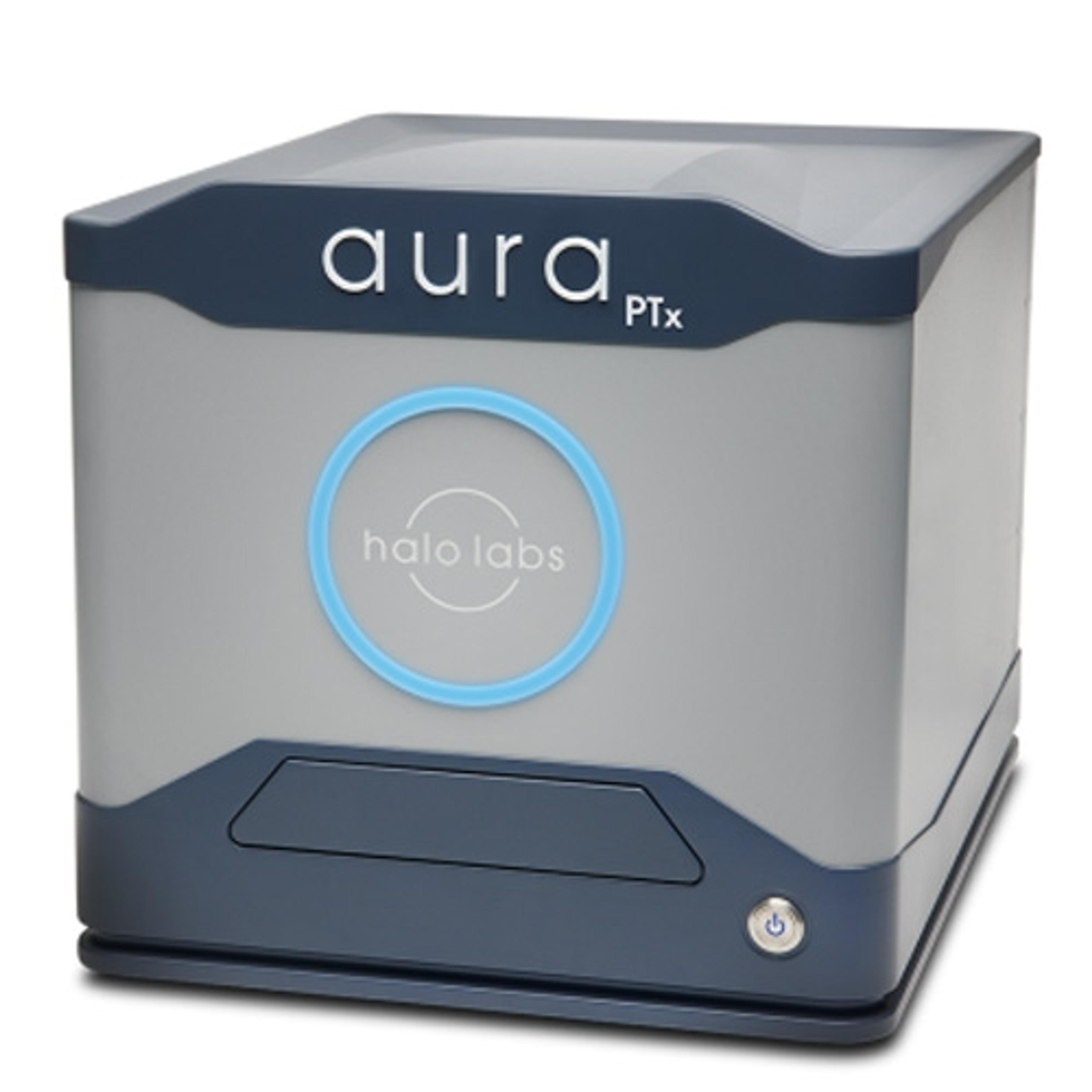
Aura PTx
Halo LabsAura® PTx gets you to your best formulation faster! Backgrounded Imaging (BMI) technology to give you accurate count, size, and morphology information about particles in your sample. Couple that with Fluorescence Membrane Microscopy (FMM), which gives you definitive ID of protein aggregates and an easy way to monitor polysorbate degradation.
Antibody development
BBI SolutionsOur unique combination of custom antibody development, lateral flow development and manufacturing services, signal enhancement technology and our mobile diagnostic platform are shaping the future of lateral flow. Our partnership approach allows you to work with one company to reach your assay goals.
Lateral flow assay development
BBI SolutionsDiscover our breadth of experience and be reassured that our Lateral Flow Development Services will deliver the best outcome for your assay.