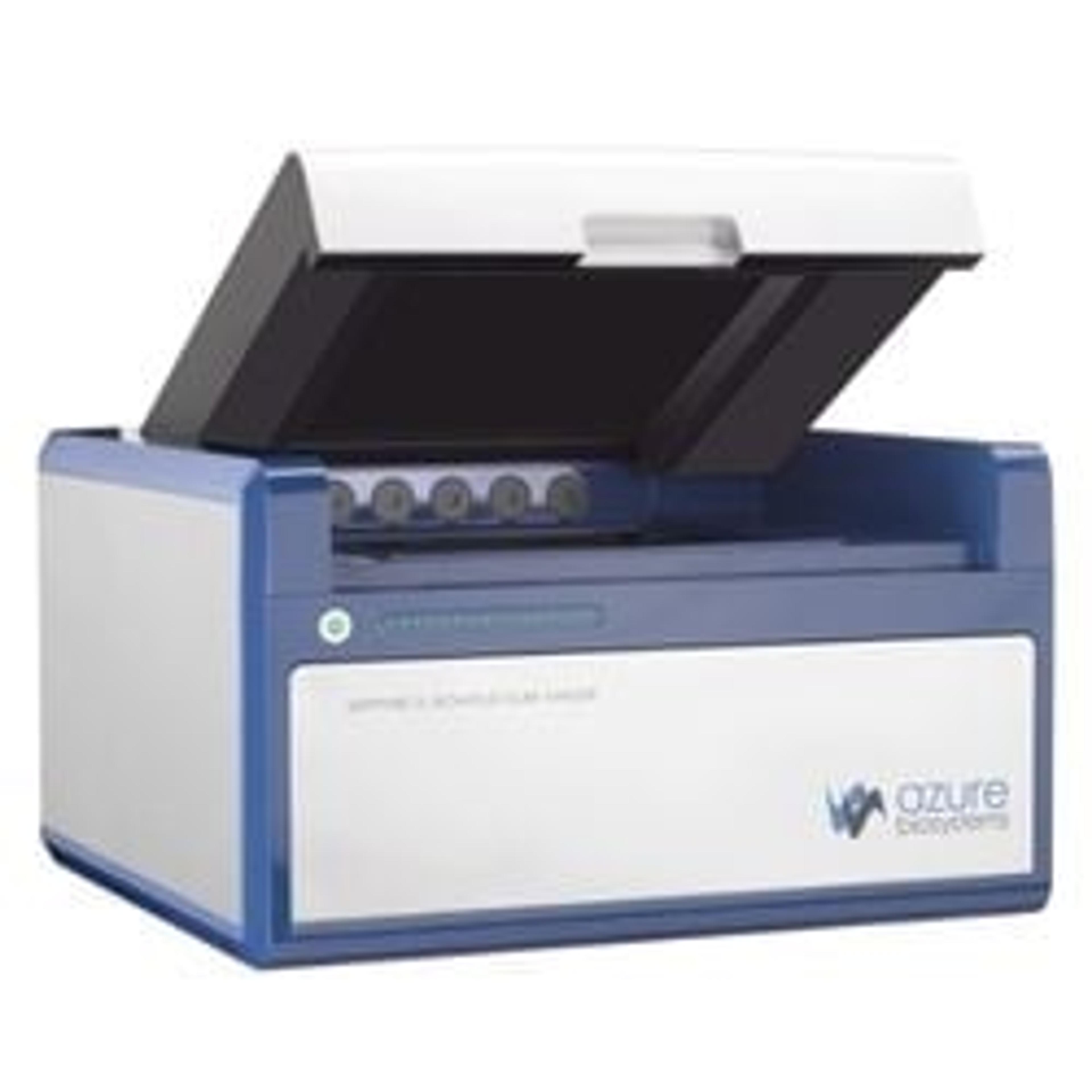
Fluorescent IHC imaging of tumor tissue arrays on the Sapphire FL
3 Jul 2024Tissue microarrays offer a high-throughput approach to histology, immunohistochemistry (IHC), fluorescence in situ hybridization (FISH), and other experiments carried out on tissue sections. They are particularly valuable for studying tumors and cancer biology, as they facilitate comparisons among different sample types. Tissue microarrays are typically imaged under a microscope, one sample at a time. Azure Biosystems describes how tissue microarrays were stained with fluorescent stains or probed with antibodies in a two-color IHC experiment, and the Sapphire™ FL Biomolecular Imager was used to capture images of the entire array. These whole-slide images can be used to catalog slides and provide an initial survey of staining success before proceeding with higher-resolution imaging.

