ResourceLife Sciences
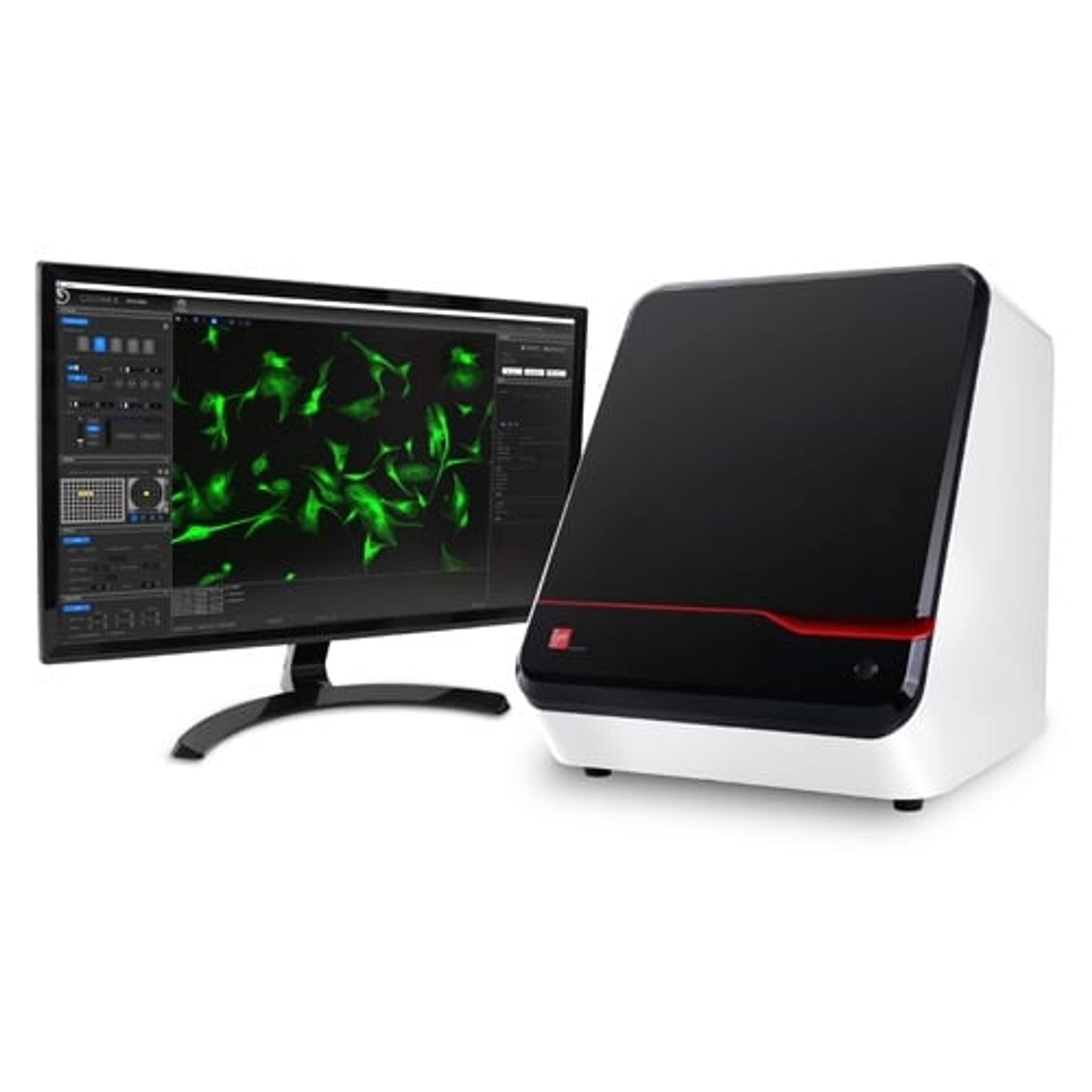
Imaging and analysis of 3D cell models using the CELENA X High Content Imaging System
24 Jul 2025Two-dimensional (2D) cell cultures have long been used in research due to their simplicity and low cost. However, three-dimensional (3D) cell models offer a more physiologically relevant environment, better mimicking in vivo conditions. Organoids, derived from human stem cells, replicate the structure and function of human organs, while 3D spheroids reflect nutrient and oxygen gradients similar to those in tissues. These models are particularly valuable in cancer research. This article outlines how organoid viability and spheroid growth can be imaged and analyzed using the CELENA® X High Content Imaging System and Cell Analyzer software.