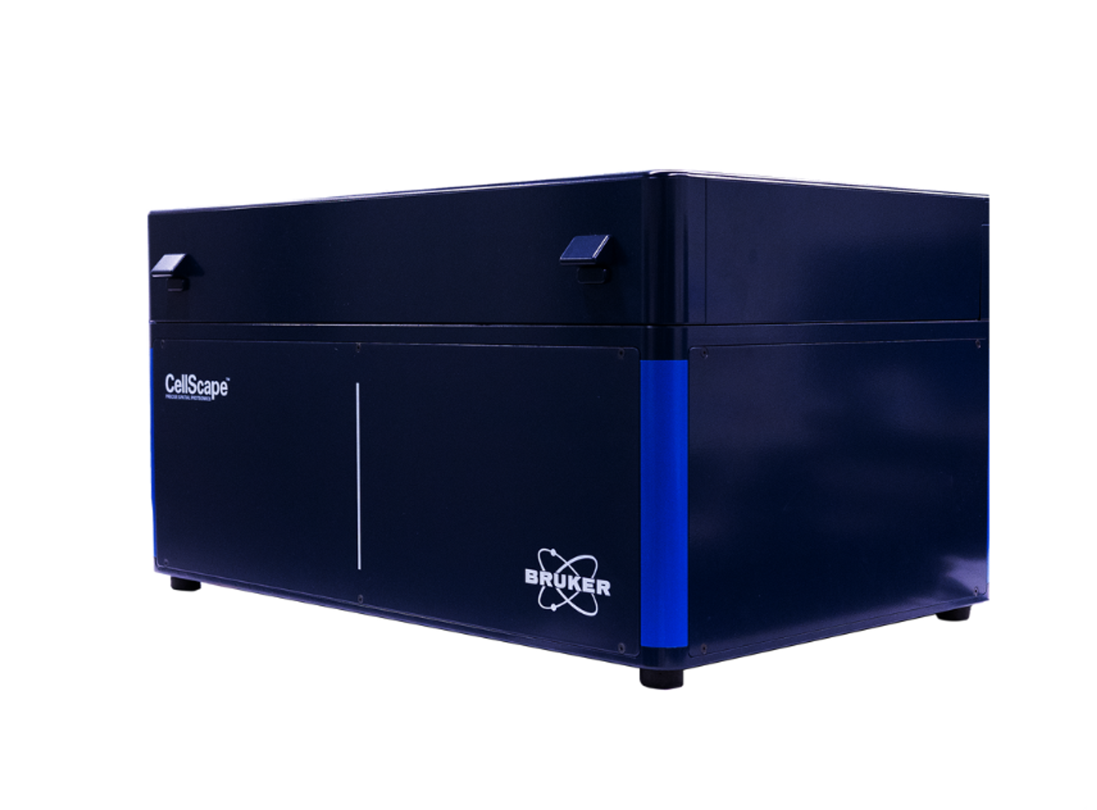
Precise spatial multiplexing of protein biomarkers for immune profiling in tissue samples with ChipCytometry
6 Jun 2023Immunohistochemistry is the most widely used diagnostic technique in tissue pathology. However, IHC is associated with several limitations including the labeling of just a few markers per tissue section and limited quantification of cell populations. As a result of plex limitations, key insights about tumor biology are missed, which could be important for advancing our understanding of tumor biology and ultimately improving patient outcomes. ChipCytometry™ is a novel image-based platform for precise spatial multiplexing that addresses these challenges by combining iterative immuno-fluorescent staining with high dynamic range imaging to facilitate quantitative phenotyping with single-cell resolution.
In this research poster, Canopy Biosciences demonstrates how standard flow cytometry standard (FCS) files are generated from multichannel OME-TIFF images, enabling the identification of cellular phenotypes via flow cytometry-like hierarchical gating. Quantification of results reveals precise expression levels for each marker in the assay in each individual cell in the sample, while maintaining spatial information about each cell.

