ResourceLife Sciences
The Art and Science of Experimental Design with Fluorophores
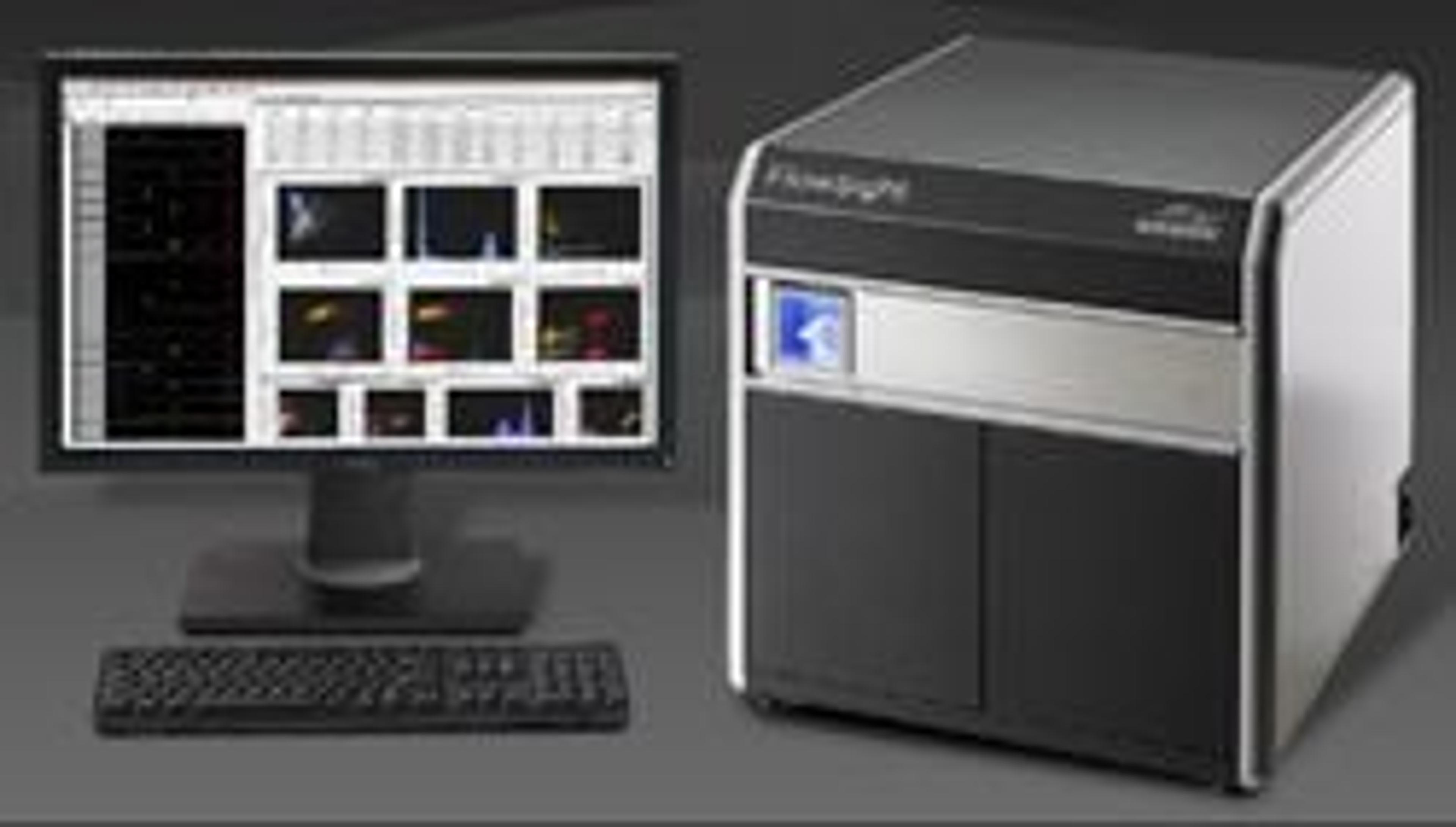
23 May 2017Imaging cytometers are at the confluence of advances in flow cytometry and light microscopy. The ability to image tens of thousands of fluorescently labeled cells results in data that are both visually striking and highly quantitative. In this infographic, learn how Amnis® technology combines lasers and optics to accommodate hundreds of fluorophores and fluorescent reagents. Build an expert palette for your own memorable discoveries with imaging flow cytometry.


