ResourceLife Sciences
Using Color Brightfield Imaging with the Cytation™ 5 to Image Hematoxylin and Eosin Stained Tissue
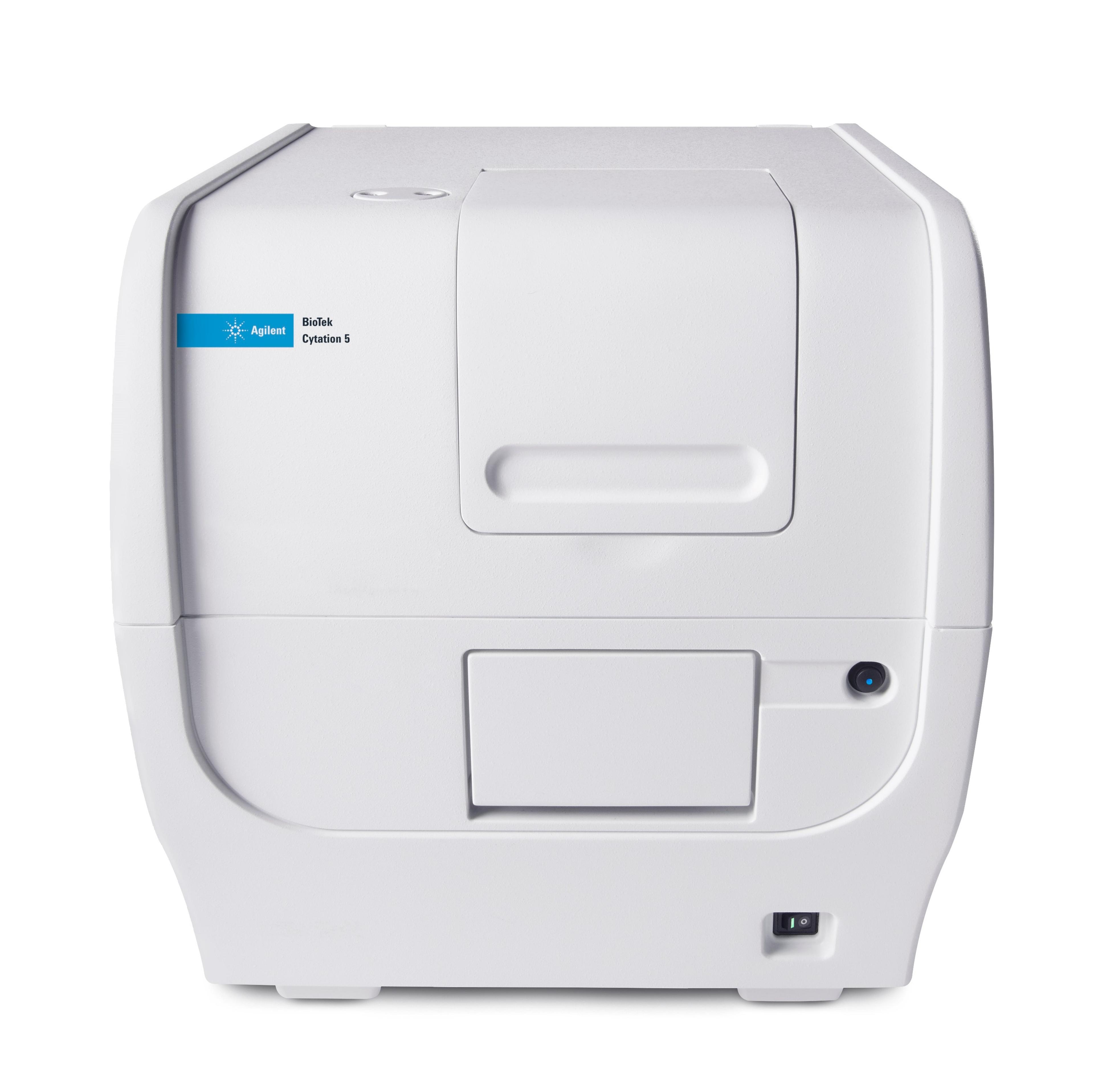
19 Nov 2014The imaging and analysis of labeled fixed and chromogenically stained cells has traditionally been accomplished using manual examination of microscopic slides with a multi-objective staged microscope. With the advent of digital color brightfield imagery, samples can be automatically imaged and the data stored for examination independent of the microscope. This application note describes the use of the Cytation 5, a low cost high-value combination cell imager and microplate reader, to rapidly image Hematoxylin and Eosin stained tissue slides.


