Cellular Pathology Community Round-Up
Latest SelectScience® news and downloads related to cellular pathology
28 Mar 2016

Latest cellular pathology news and application article downloads from the SelectScience® Library
In the news recently, learn why ’de novo’ FDA clearance of whole slide imaging devices is significant, find out how a new device could diagnose cancer rapidly at the cellular level, and discover more about scanning near-field optical microscopy.
1. Article: Elephants in the Room: Whole Slide Imaging Devices
In mid-January the Digital Pathology Association (“DPA”) announced that the ‘de novo’ application process may be acceptable by the FDA for clearance of whole slide imaging devices (“WSI” devices) for primary diagnostic uses. Why is this significant? The de novo process lowers the primary hurdle for marketing approval from that of a class III device to essentially a class II device.
2. News: Tews Laboratory use LaVision BioTec Technology for Tumor Study
LaVision BioTec, developers of advanced microscopy solutions for the life sciences, report on the work in the Tews Laboratory which is studying the molecular mechanisms of tumor invasion using an UltraMicroscope for enhanced imaging of cells.
A University of Texas at Arlington electrical engineer has developed a novel cancer cell detection method that will improve early diagnosis through a tool that tracks cellular behavior in real time using nanotextured walls that mimic layers of body tissue.
Dako, an Agilent Technologies company and a worldwide provider of cancer diagnostics, has announced that the FDA has approved the expansion of the intended use of Dako PD-L1 IHC 28-8 pharmDx to include patients with melanoma.
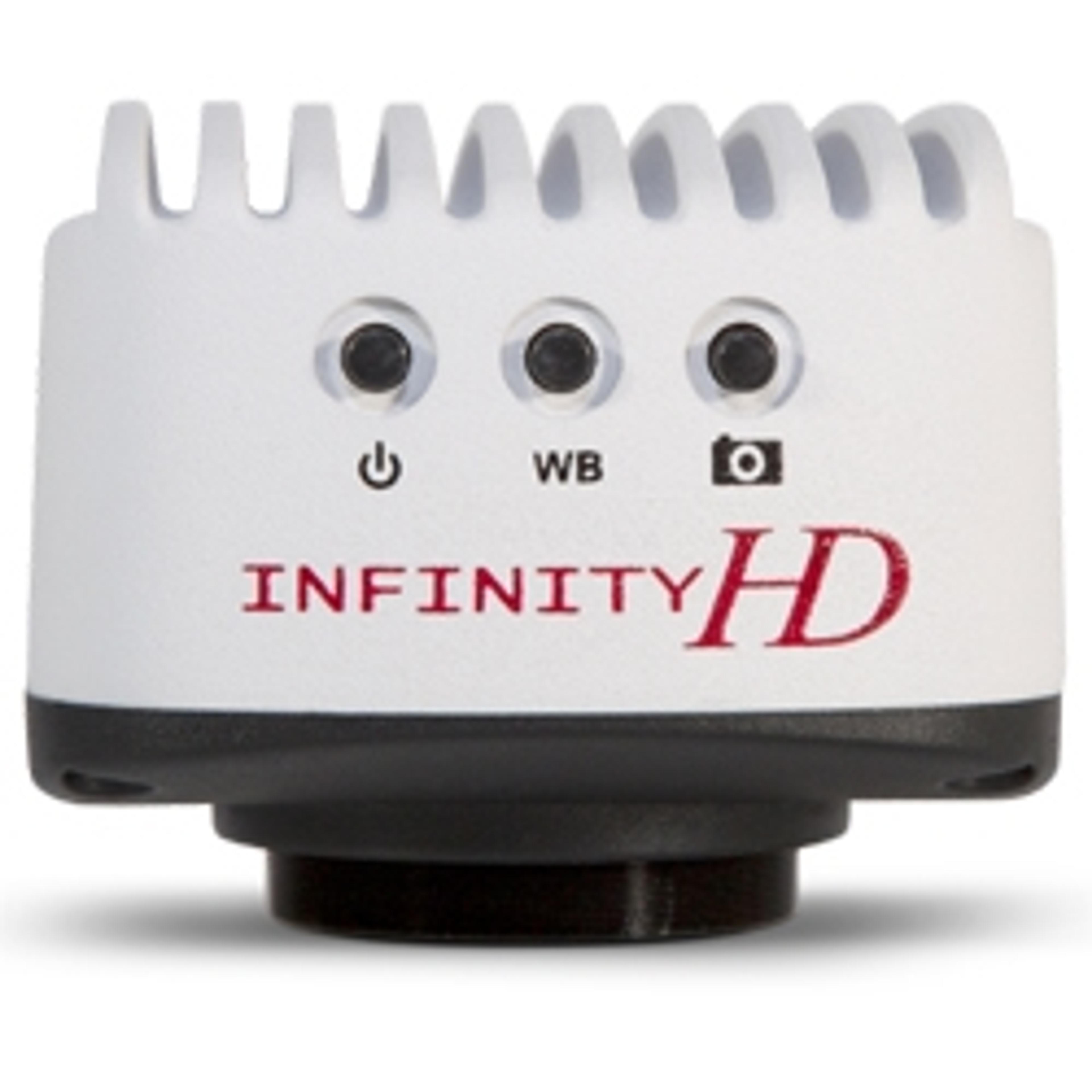
In this case study Lumenera demonstrate the high image quality of its microscopy cameras, image quality, especially clarity of color, is essential in dermatopathology as the diagnosis relies of microscopy photographs of stained tissue. Lumenera’s INFINITY 2-5 CCD camera was suited for the teaching side of the dermatopathology clinic as its ImageJ application allowed for easy editing and viewing on the Mac system.
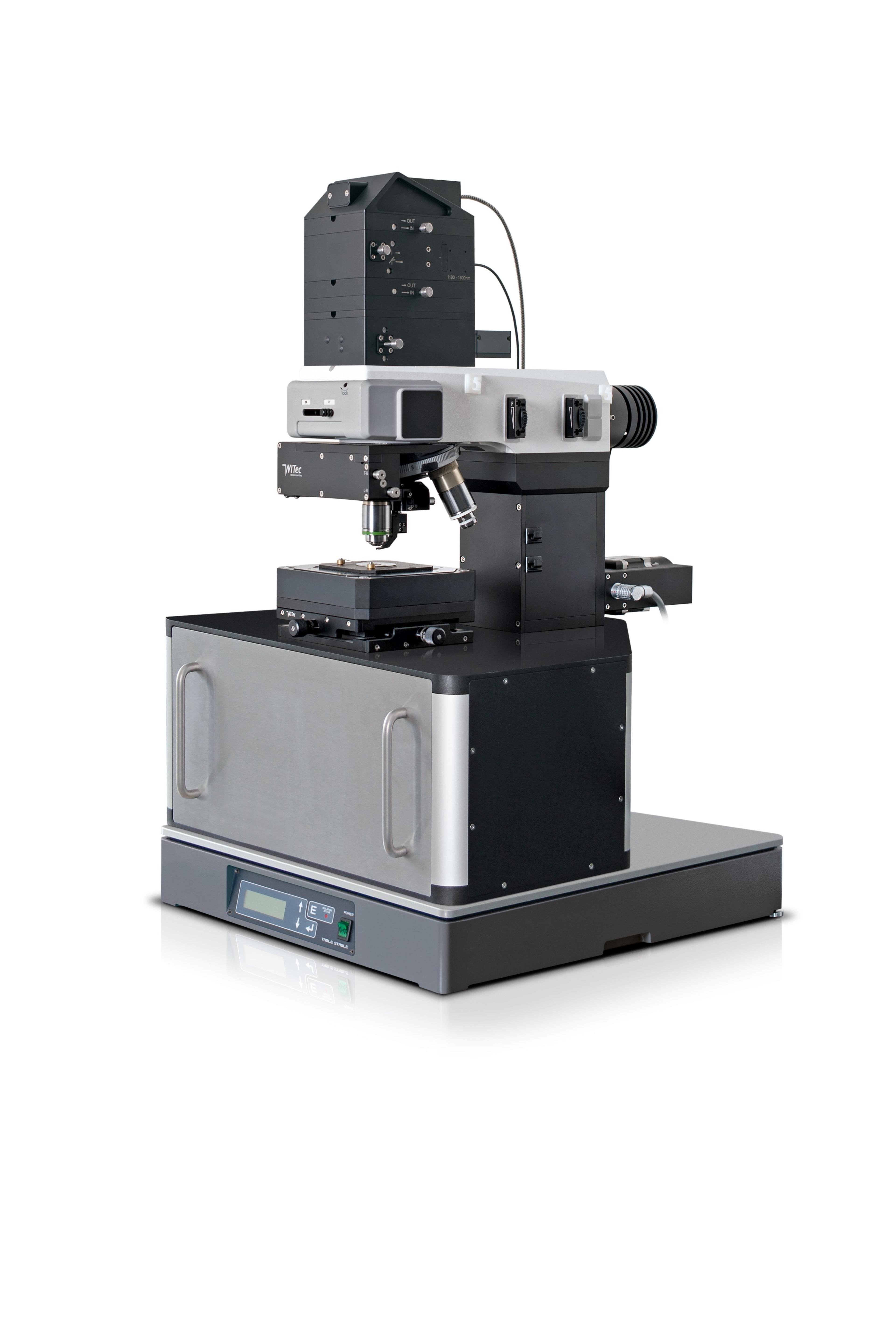
6. Application Download: Scanning Near-Field Optical Microscopy - Histological Microtome Cuts
Scanning Near-field Optical microscopy allows tissue to be viewed in air or liquid at high resolution without taking up time with sample preparation. This application note from WITec GmbH uses a variety of rat organs to show the results of Scanning Near-field Optical microscopy, showing the different structures that can be clearly seen in the tissue after staining.
This application from Waters Corporation demonstrates that desorption electrospray ionization (DESI) imaging can be utilized as a non-destructive imaging technique. This allows multiple analyses on the same tissue section at different spatial resolution.
Image: anyaivanova/Shutterstock