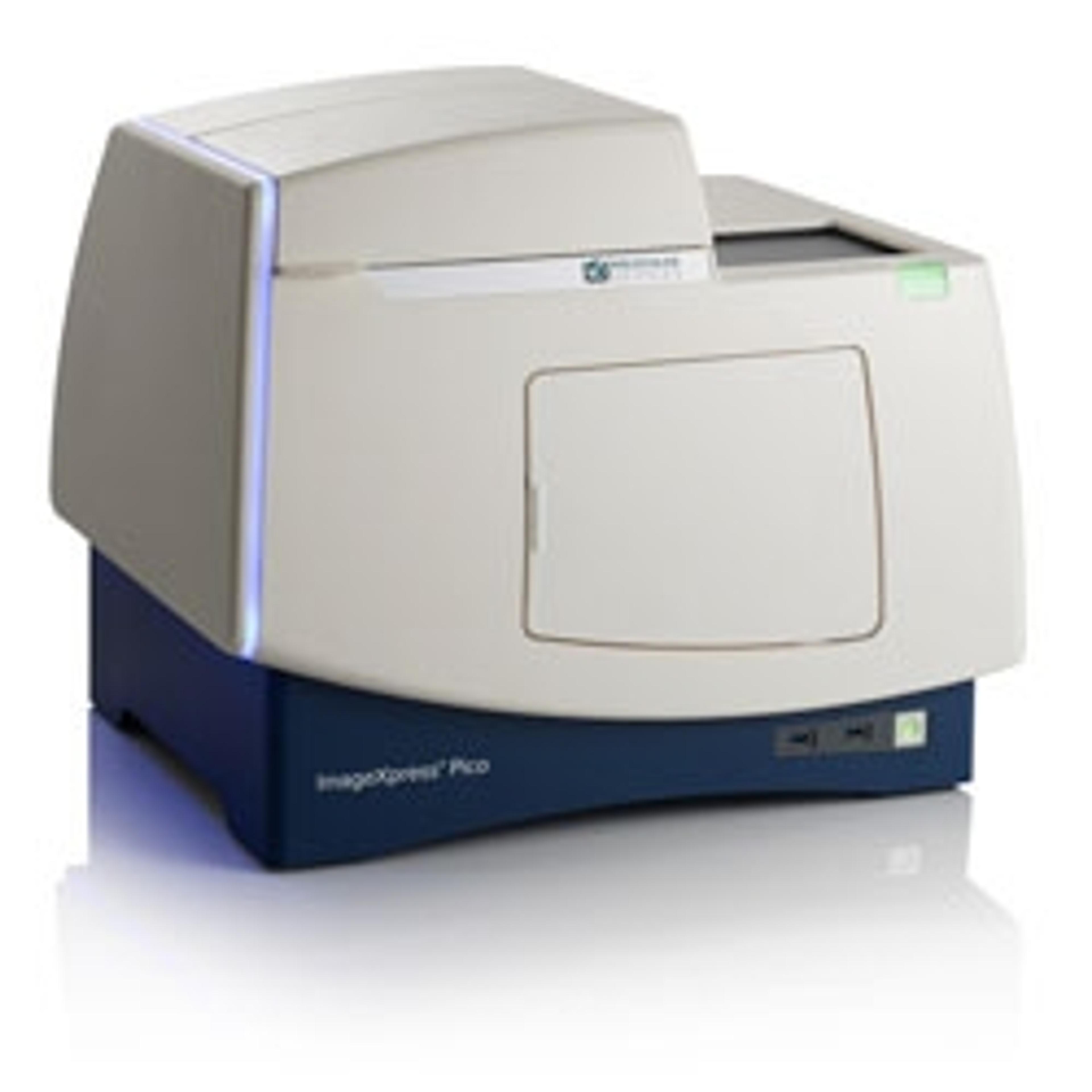
Everything you need to know about the ImageXpress Pico Automated Cell Imaging System
We put your most pressing questions to the team behind Molecular Devices latest cellular imaging and analysis system to find out how it could make an impact in your lab
11 Mar 2020
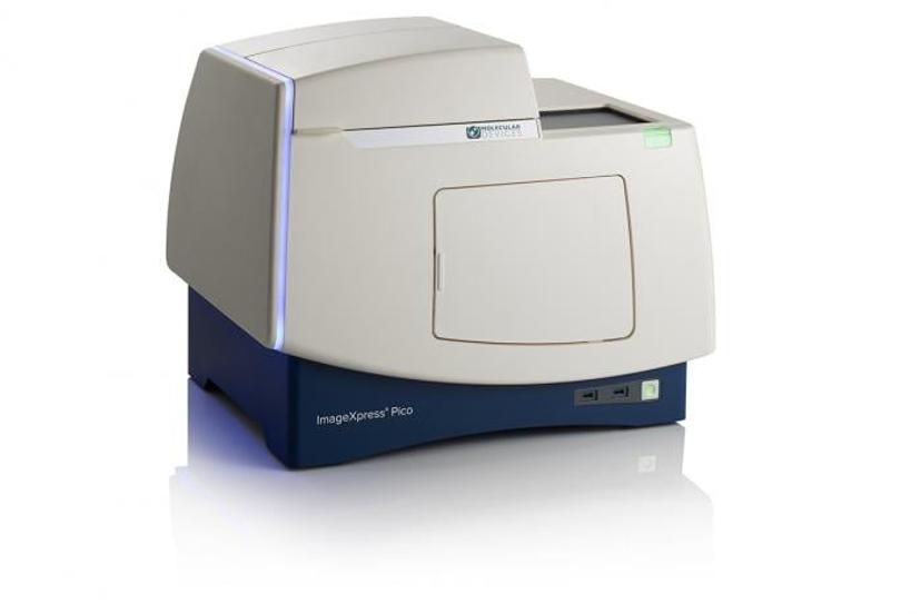
Depending on your application and exactly what you are researching, there are multiple imaging techniques available, ranging from confocal microscopy to electron microscopy and more - each has its own benefits and challenges. In this FAQ article, we take a closer look at the ImageXpress Pico Automated Cell Imaging System from Molecular Devices. Find out how this compact, fully automated imaging system with integrated CellReporterXpress software is designed to reduce the need for conventional, tedious manual microscopes that may be prone to error. The multiple imaging modes, with objectives ranging from 4x to 63x, can operate in colorimetric, brightfield, fluorescence, or Digital Confocal* 2D on-the-fly deconvolution.
How can the ImageXpress Pico Automated Cell Imaging System be used for cell counting?
Compact system with intelligent image acquisition and analysis
Molecular Devices
Cell counting is a technique used across numerous biological experiments. Commonly in cell-based assays, such as drug compound toxicity or cell proliferation, there’s a need to assess the number or density of cells in a well. Automated imaging can speed up the process while reducing manual labor and human error. Cells can also be counted using a variety of methods, for example, the detection of nuclear dye with fluorescent imaging or label-free cell counting under transmitted light. Read how the imaging system is being used for multi-parametric assessment of cell phenotypes>>
How can I image cell death assays, for example, for apoptosis analysis?
Autophagy vs Apoptosis
Autophagy - degrades internal structures in its own cytoplasmic material. These structures are degraded using lysosomes.
Apoptosis - the cell labels itself for cell death. The cell breaks down into smaller fragments inhibiting its function or ability to replicate
Cells can die by different mechanisms. The two common methods are apoptosis, also known as programmed cell death, or autophagy. These play an important role in multiple biological processes, from embryonic development to normal tissue and immune system maintenance.
Disruption in the normal regulation of apoptosis and the presence of autophagy are both thought to be markers of diseases such as neurodegenerative diseases and cancers.
Characteristic changes in cell morphology occurring during these processes — such as cell shrinkage, nuclear fragmentation, chromatin condensation and mRNA decay — can be imaged during both autophagy and apoptosis.
- Find out more about detecting autophagy using imaging systems>>
- How analysis of apoptosis is imaged>>
- Cytotoxicity assessment using imaging of live/dead assays>>
How can the ImageXpress Pico Automated Cell Imaging System be used for angiogenesis research?
Angiogenesis, the formation of new blood vessels, has recently become a focus in cancer therapeutics. Imaging techniques can help advance this research by visualizing the process and enabling the development of 3D tissue models. An expert in the field Dr. Bas Trietsch, Chief Technology Officer at MIMETAS, is trying to overcome conventional tissue culture pitfalls when using animal models. We interviewed him at SLAS Europe 2019 to discuss imaging developments in this field.
Read this article to find out more>>
Are there configurable options to support multiple assays?
With a wide range of viable assays for scientists to choose from, having an instrument that can adapt to the needs of each assay is often sought after. The ImageXpress Pico Automated Cell Imaging System has over 25 preconfigured templates available, ranging from simple cell counting to neurite tracing.
Find out how to image neurite outgrowth>>
Every experiment has an end goal of generating publication-ready data. This system can generate results that can be visualized in various formats including heat maps, scatter plots, tables, bar charts, and movies ready to be included in your publication.
How can I significantly increase the resolution of my images?
Assays that require precise identification of subcellular structures or molecules within different cellular compartments can be difficult to identify. The ImageXpress Pico’s optional Digital Confocal* 2D deconvolution algorithm helps reverse optical distortion on-the-fly, allowing you to quickly capture images with higher signal-to-noise for more precise segmentation and analysis.
Discover more of the latest resources in cell imaging>>
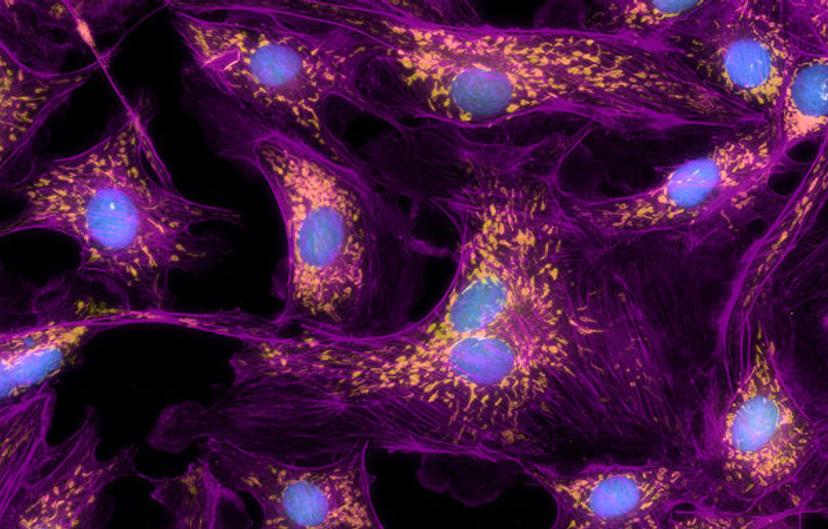
40X Digital Confocal* images of BPAE cells with nuclei (blue), mitochondria (pseudocolored yellow), and actin (pseudocolored purple) staining. Images were captured on the ImageXpress Pico image courtesy of Molecular Devices
How can the ImageXpress Pico system help reduce the burden for core facilities?
Many core facilities have high-throughput imaging systems running day and night for complex screening assays. The ImageXpress Pico is helping to alleviate the user burden from these high-end systems by offering walk-up ease-of use for lower throughput and less advanced imaging assays. Rather than spending time learning how to use a system whose full capabilities are not taken advantage of, scientists can get started with the ImageXpress Pico with very little training, thanks to the intuitive software interface. The system’s lab-friendly price also allows researchers to afford the convenience of automated imaging and analysis on their own lab bench, further alleviating the demand on core facilities.
How does the live preview in the CellReporterXpress software work?
Click-to-center functionality enables users to visualize their sample prior to data acquisition using the virtual joystick to pan around and interactively adjust the focus, exposure and position. This makes establishing acquisition settings much easier.
Data can be visualized at multiple levels, from plate overview to individual cells. A range of data visualization tools empowers users to learn as much as possible from their images and assays enabling them to verify and have confidence in their data.
These new features enable scientists to expand their research to include more relevant cell model systems and more complex analysis at an affordable price point
Jeff McMillan Senior Imaging Product Manager at Molecular Devices

What is the benefit of environmental control and z-stack acquisition?
Cells are sensitive to their environments. The environmental control feature of the ImageXpress Pico allows the user to perform long-term live-cell experiments while maintaining optimal conditions for the experiment. Multi-day, time-lapse, and live-cell assays can be run using the onboard environmental system with options for temperature, humidity, CO2, and O2 control. Environmental conditions are monitored and recorded throughout each timelapse experiment, allowing the user to verify these external factors in the event of abnormal data points.
Discover how cell migration can be measured using discontinuous time-lapse imaging of live cells>>
Cells are complex 3D structures and information can get lost amid the layers. The Z-stack acquisition feature uses CellReporterXpress Automated Imaging and Analysis Software to acquire a series of images at different focal points, capturing more detail than with a single slice. Users can include all slices or select which slices to include in the final projection. This can build a bigger picture of the cell or allow isolated pictures where necessary.
Do you use the ImageXpress Pico Automated Cell Imaging System in your lab? Write a review today for your chance to win a $400 Amazon gift card>>
*ImageXpress Pico Digital Confocal uses AutoQuant 2D Real Time Deconvolution