Masterclass in modern microspectroscopy
In this on-demand webinar, Dr. Michael Bradley describes how to draw big information from tiny samples using microspectroscopy
7 Mar 2022

Pressure to deliver successful results faster seems to increase all the time. However, using state-of-the-art infrared (IR) microscopy tools can help meet these demands. Microspectroscopy can analyze particulates, fibers, inks, contaminated surfaces, and more, making it a workhorse tool across a wide range of industries, from environmental to automotive to pharmaceuticals. Forensics and research labs need the capability to address a broad range of sample types.
In this expert SelectScience® webinar, now available on demand, Dr. Michael Bradley, product manager of FTIR spectrometers and microscopes at Thermo Fisher Scientific presents how you can improve the speed and accuracy of your microspectroscopy results.
Watch on demandRead on for highlights from the Q&A discussion and register now to watch the webinar on-demand
What's the smallest size and thickness that FTIR can analyze, and can you share what the best setting is for this type of sample condition?
MB: The wrapped IR microscope is good to below five microns without using ATR, so it offers a very high spatial resolution. As far as the thickness goes, that will depend very much upon the sample. We try to keep them down to a few microns if you're doing transmission because the thicker they are, the more absorption you get, and the bands flatten out and you get totally absorbing bands, things like that.
As far as infrared reflection absorption, that has the problem where you're going through the sample twice, so the problem is somewhat compounded. But still, as I said, I'm looking at layers within that laminate I showed you that were down to below 10 microns thick, and those were done on a laminate that was roughly 15 microns thick. It was cut with a razor blade, not a microtome. So, it really depends on the sample, but those are some rough numbers of what we work with.
Reflection versus ATR, how deep does the FTIR penetrate to give information?
MB: The penetration depth depends upon a lot of parameters, there's an equation that talks about that. For ATR, we use a germanium crystal, which gives you a very shallow depth of penetration, so you're probably less than half a micron deep into the sample and you're very much looking at the surface phenomenon. Like with the currency, you're looking at the ink on the surface. Frequently, you may not even see down below the ink layer, but it just depends upon the penetration.
For the reflection absorption and the reflection types of experiments, that depends again upon the material, because the IR beam will go as deep as it can into the sample, which can be several microns. It really depends upon the absorptivity of the sample itself. That's a somewhat difficult question to answer, but with the ATR you have good control over that.
What are the advantages of having a tandem microscope and bench, as opposed to just a standalone microscope, if your samples are only for microscopy?
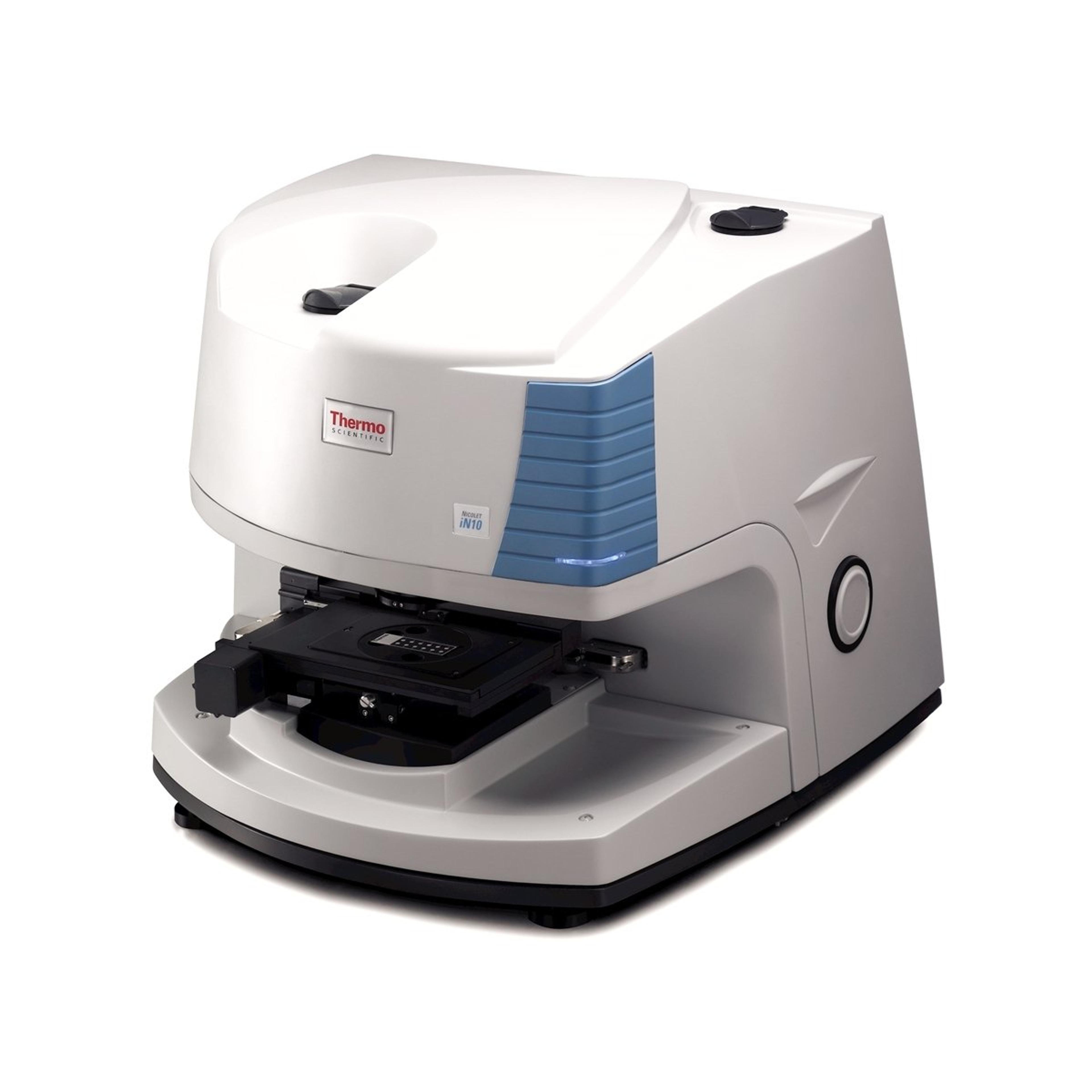
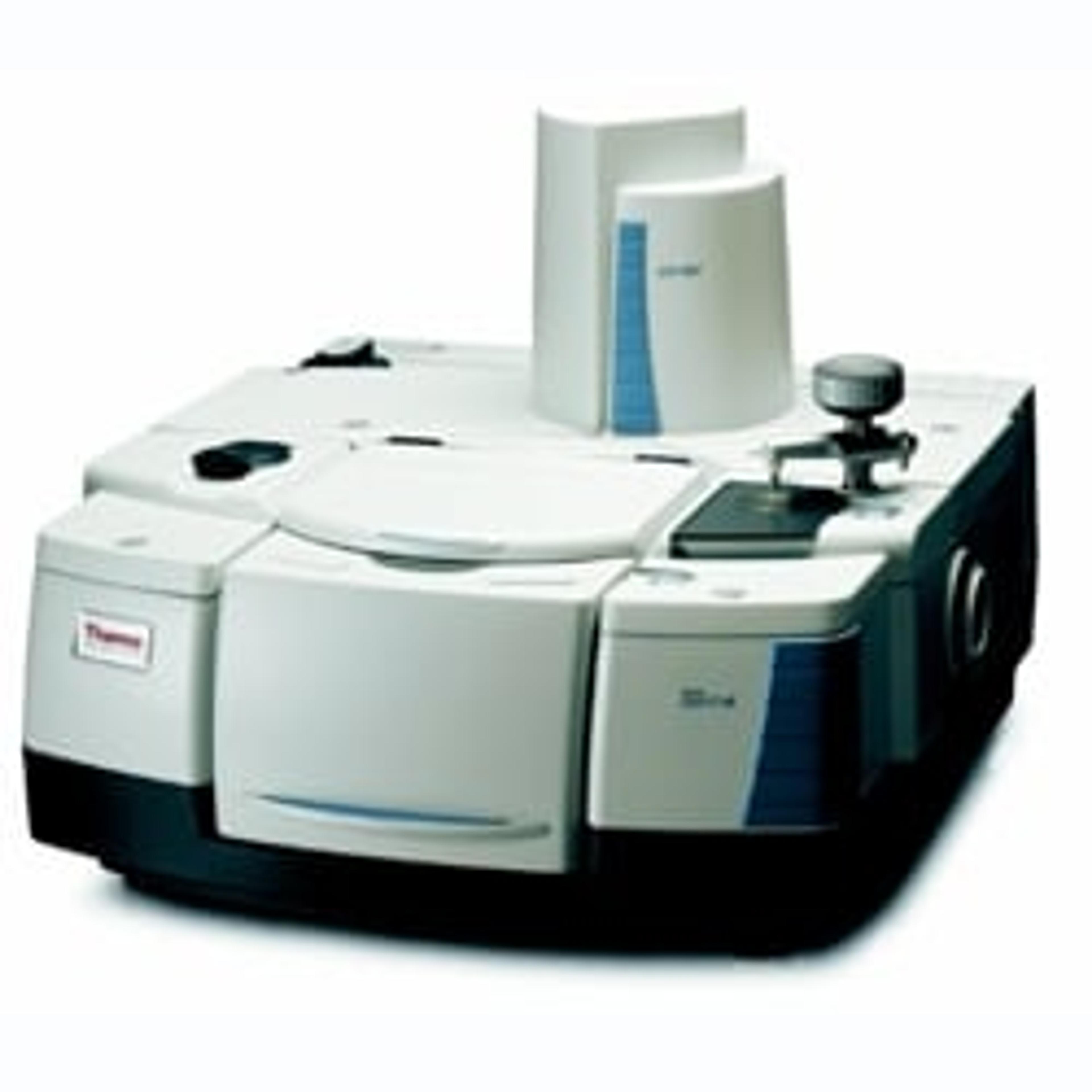
MB: If your samples are only for more microscopy, that's why we offer the standalone instrument, the Nicolet IN10. However, with the wrapped IR and the attached Nicolet IS50 that you have on the front end, you get several other capabilities, including Fourier transform Raman, thermogravimetric analysis, bulk ATR - all the tools that you would expect out of a research-grade spectrometer. Also, in the long run, you will be able to pick up various spectral ranges, because the spectrometer can do that, and gives us the ability to control some other parameters.
But the primary advantage here is the ability to connect to it and get all those other capabilities. If you don't need them, then that would be looking at the IN10. Really, you need to sit down with the salespeople and the applications people to look at your app and decide which one is the best route for you to go.
What are the developments required for microparticles that are only microns in size?
MB: Thermo Fisher Scientific is a very broad company, it has a lot of expertise, and among that is an imaging technology that was heavily used in electron microscopy. You may be familiar that FEI is now part of Thermo, and they developed a lot of 2D and 3D imaging technologies to work with that. So, what we did was we imported that technology into the FTIR microscope to give us the most efficient and effective way of detecting the particles.
The other thing that was developed for this tool was the path that the spectrometer takes through the sample is optimized. We don't just start in the upper left-hand corner and go linearly through the sample, that would require changing the aperture at each point or could require that. Instead, what we do is we've set one aperture, and then we collect all the particles that use that aperture.
By using the visual tools to optimize the data collection, and then optimize the path, we're giving you a very high-speed analysis without the need for an imaging tool or something like a quantum cascade laser. It's just not necessary for this kind of analysis.
How does the wrapped IR scope differ from the continuum scope? What upgrades does the wrapped IR scope offer?
MB: It's a much more modern microscope and has a much higher spatial resolution and a much higher quality camera on it. This is a full, large-scale, high-resolution technology, enabling resolution down to below a micron for the visible spectrum. And as I said, you're below five for the infrared, that's the primary change.
The other massive change is the import of the paradigm software, which is full 64-bit. I recently collected a visual image that was eight gigabytes in size, something that the previous generations of software could not have handled. The paradigm software allows me to handle that and then do the data collection and the analysis.
What we're looking at is an entirely new user interface, as well as an entirely new technological platform. This is a completely different platform. Even if some of the components look the same, they're not. The other thing is that you can use thick samples. With the continuum, you're limited on how thick the samples could be that you can mount. As the images showed, you can mount books, you can mount paintings, and other things on the wrapped IR microscope platform.
To learn more about microspectroscopy, watch the full webinar here>>
SelectScience runs 10+ webinars a month across various scientific topics, discover more of our upcoming webinars>>