Single-cell genomics at the Memorial Sloan Kettering Cancer Center: Developing innovative methods
Dr. Ronan Chaligné, Head of the Single-Cell Analytics Innovation Laboratory, discusses advances in single-cell methods advancing cancer research
2 May 2022
Single-cell genomics is transforming our understanding of complex biological questions. The latest technologies enable the transcriptome to be analyzed in millions of cells simultaneously. This provides significant insight into individual cellular differences and plays an important role in cancer research where cells are heterogeneous within the same tumor, making bulk analysis difficult. With a broad range of samples to analyze, there is a demand to improve and streamline methodology so that maximum information can be obtained from each experiment.
Here, we speak with Dr. Ronan Chaligné, Head of the Single-cell Analytics Innovation Laboratory at Memorial Sloan Kettering Cancer Center (MSK), one of the largest cancer centers in the world, about the studies his lab performs and how new sample preparation technology has impacted single-cell genomics workflows.
Advancing single-cell methods
The Single-cell Analytics Innovation Laboratory (SAIL) has two goals; to produce high-quality single-cell data for research at MSK, and to develop and optimize new single-cell methods that will help answer new biological questions.
Chaligné, who began his research career at Institut Curie studying epigenetic instability in breast cancer, followed by Weill Cornell where he investigated DNA methylation instability in blood cancers, is passionate about method development. At SAIL, Chaligné manages a team of 10 scientists split between bench-side and computational research. The lab works on single-cell genomics and collaborates with over 60 research laboratories a year. While many samples come from cancer patients, the team also works on fundamental biological questions. “As one of the biggest cancer centers in the world, we work with a lot of clinical samples from patients, from all varieties of cancer, but there is also a strong focus on fundamental science,” Chaligné shares. “We work with Drosophila, zebrafish, mouse models, organoid models, and patient-derived xenograft models. So, we really are working with a variety of samples, and trying to apply new single-cell genomics methods to all of them, to try and answer the biological questions of our collaborators.”
Challenges in single-cell genomics
Working across many sample types, the team at SAIL faces several challenges as they develop single-cell methods, both at the bench side and in data analysis. One concern is limited sample volume. “We can work on really tiny pieces of biological material, and we are trying to extract as much information as we can, which is of course challenging,” Chaligné says. “We are always trying to prepare the sample without any outside nucleic acid or contaminants coming into the experiment.”
In addition, many of the samples sent to SAIL are very fragile and must be handled carefully to ensure their cellular composition and transcriptional states are not altered. Working with clinical surgery samples also means the lab can face logistical challenges if a patient’s surgery is delayed or postponed.
From the computational side, data is often incomplete. “You never capture 100% of the information from a single cell. So, computational biology is used to apply mathematical modeling to try to impute missing biological information from them,” Chaligné explains.
Applying new cell separation technology
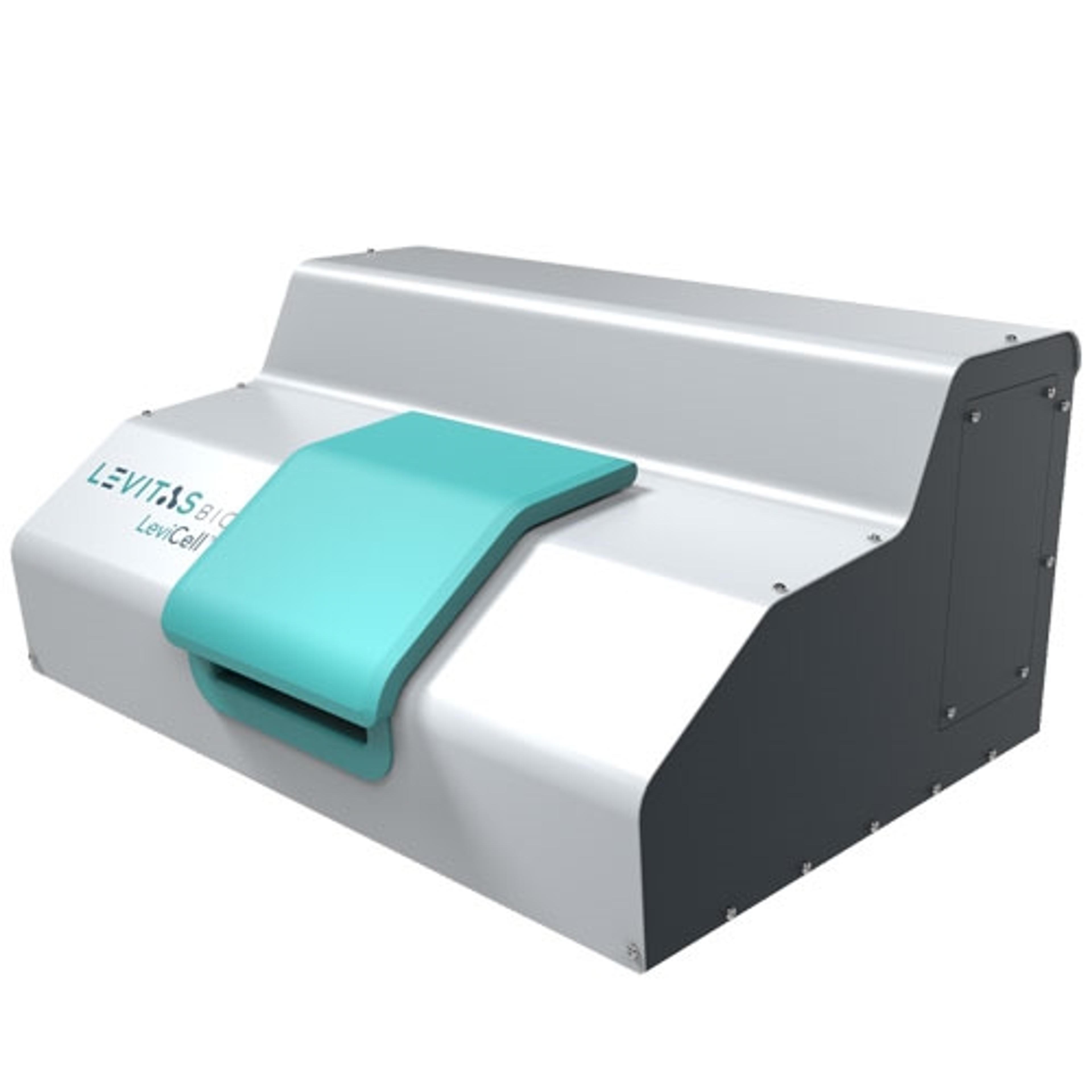
SAIL has recently implemented the LeviCell™ system (LeviCell) from LevitasBio, which utilizes a simplified workflow for label-free cell enrichment and purification – something which has helped the Chaligné SAIL team to overcome some of the challenges around fragile sample preparation.
Traditionally single-cell sorting is performed using fluorescence-activated cell sorting (FACS). However, while this is suitable for many experiments, there are some limitations to this technique, especially when working with delicate cells. “The pressure the cells are facing in FACS sorting is around 10 psi, which is pretty high, and some cells are really sensitive to that, and will die. Dendritic cells, for example, tend to die quickly after sorting,” says Chaligné. Similar issues are faced when working with cerebrospinal fluid samples, for example, where recovering viable cells following cell sorting is also challenging. “For those samples, LeviCell has been a game-changer because now we can quickly and gently extract live cells and remove all the debris and the dead cells. This has allowed us to apply our single-cell genomics method after using the LeviCell,” Chaligné says.
Furthermore, the LeviCell system uses magnetic levitation to precisely separate all cells in the sample to collect high yields of live cells, based on intrinsic properties, including density and magnetic susceptibility. Consequently, the proportion of live versus dead cells in the starting sample does not impact the time taken to separate viable cells. “If you have FACS sorting of a sample, and your samples are just 5% live cells, it will take you a long time to select those cells because the population of interest is small,” describes Chaligné. “In the LeviCell, because the whole sample is selected at the same time, the time that it's in the machine is constant, and you are not really impacted by the fact that your populations may be smaller or bigger. You let it sit in LeviCell for 20 minutes, and it's done.”
The LeviCell system has also helped the SAIL team overcome logistical challenges when working with surgery samples. “Usually, our collaborators want to treat clinical samples fresh, because you have better quality, you can dissociate the cells, and you can sort specific cell populations. But one of the caveats, when you do that, is that clinical samples are frequently postponed,” explains Chaligné. Delayed samples can lead to missed FACS appointments, leaving samples unable to be processed. “Now we have LeviCell. It's logistically easier for us. We can switch on the machine in two minutes, and we can just allow the clinical sample to arrive,” says Chaligné. “Being able to just load the sample, enrich for live cells, remove dead cells and debris, and have a good cell population, viable for the single-cell method is perfect.”
Chaligné has received a high standard of support from the team at LevitasBio and has even been involved in the beta testing of new products. “I'm really looking forward to everything they're trying to develop,” says Chaligné.
The future: Entering the era of multiomics
Looking to the future of single-cell analysis, Chaligné highlights the expanding techniques used in the field to gain more information from each experiment.
“We are now entering the era of multiomics. So far, we've been trying to improve gene expression profiling, to be more efficient at how many genes and transcripts we could capture per cell. Now, we are really entering the era where we are trying to capture more information than just gene expression. We are also capturing surface proteins, transcription factors, and chromatin landscape, amongst others. Thanks to methods developed by groups like Aviv Regev or Peter Van Galen, we can also capture mitochondrial DNA mutation, which allows us to perform lineage tracing,” enthuses Chaligné. “All these things we can almost capture in one experiment. I really see that as trying to squeeze as much information as possible from one single cell, so I think that is the way the field is advancing now, and that's exciting.” On top of this, spatial information capture will also play a key role in advancing single-cell analysis.
Together, these methods will help to further our understanding of heterogeneous diseases like cancer, from identifying which characteristics cause the expansion to why some clones are resistant to treatments. The single-cell analysis will also provide more insight into pre-cancer studies, where precancerous clones may be present decades before any cancer arises. Chaligné concludes: “As someone who really focuses on technologies, I like the way new technology is advancing biology and how it's helping answer new biological questions.”
Do you use LevitasBio products in your lab? Write a review today > >
Related article: Innovative cell technology enabling new discoveries for B-cell leukemia treatment