How to Optimize Your 3D Cell Culture with Organoids, Scaffolds and Hydrogels
Dr. Richard M. Eglen of Corning Life Sciences shares expertise on optimizing 3D cell culture assays with the latest technologies
13 Dec 2018
Three-dimensional (3D) cell culture is becoming well embedded in human disease research, enabling scientists to recapitulate molecular mechanisms between different cell types in a spatial context. In this webinar, SelectScience is joined by Dr. Richard M. Eglen, Vice President and General Manager of Corning Life Sciences, for a compelling live presentation on advances in 3D culture technologies. Catch the highlights from the Q&A session below or watch the webinar in full on demand.
Q: If you were to recommend a method for 3D culture, which one would be your first choice?
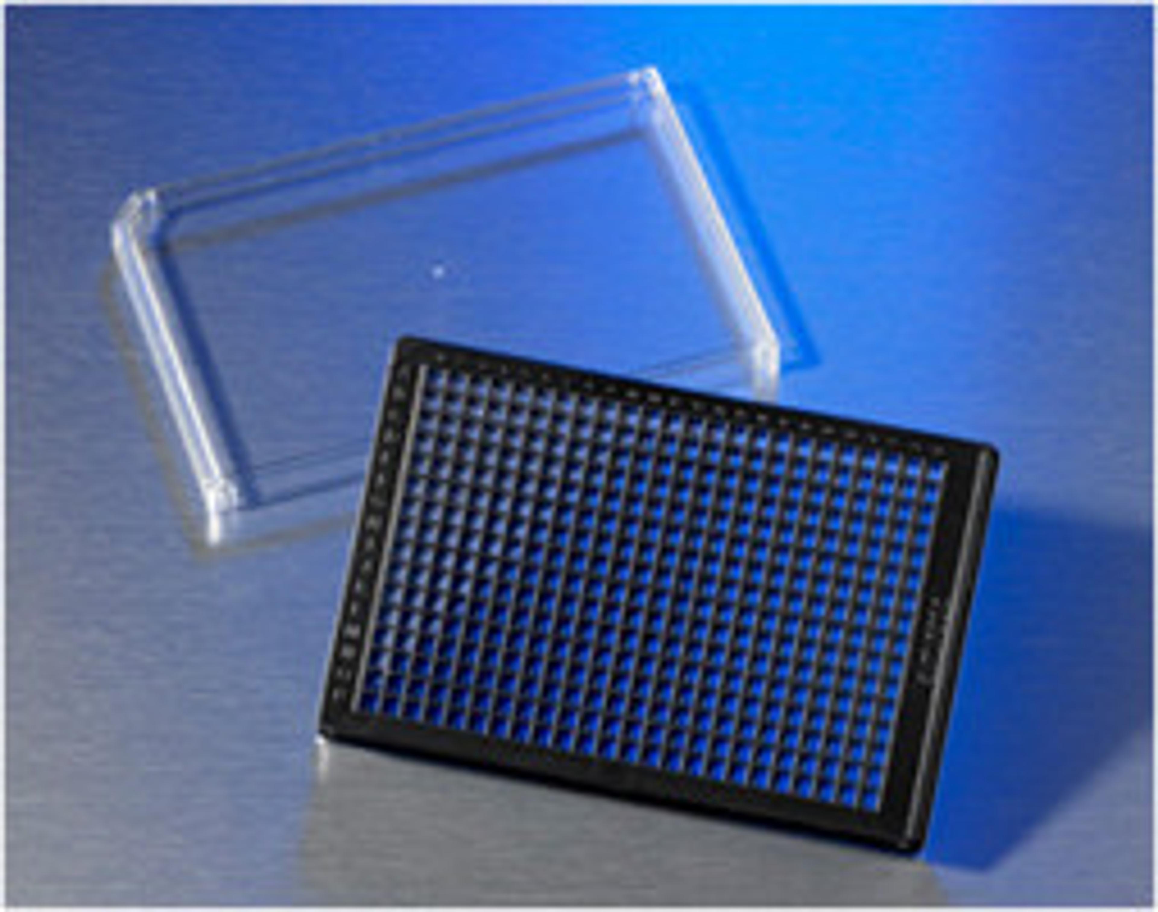
R.E: The Corning® Ultra-Low Attachment spheroid microplates are ones with which people have found a great deal of success. The fact that you can grow the cells and then do the assay in the same plate, without a separation step, is a considerable advantage.
As a first step into the field, I would recommend the spheroid microplate system – you should have good success rates with the use of spheroids and the reproducibility of the assays with this approach.
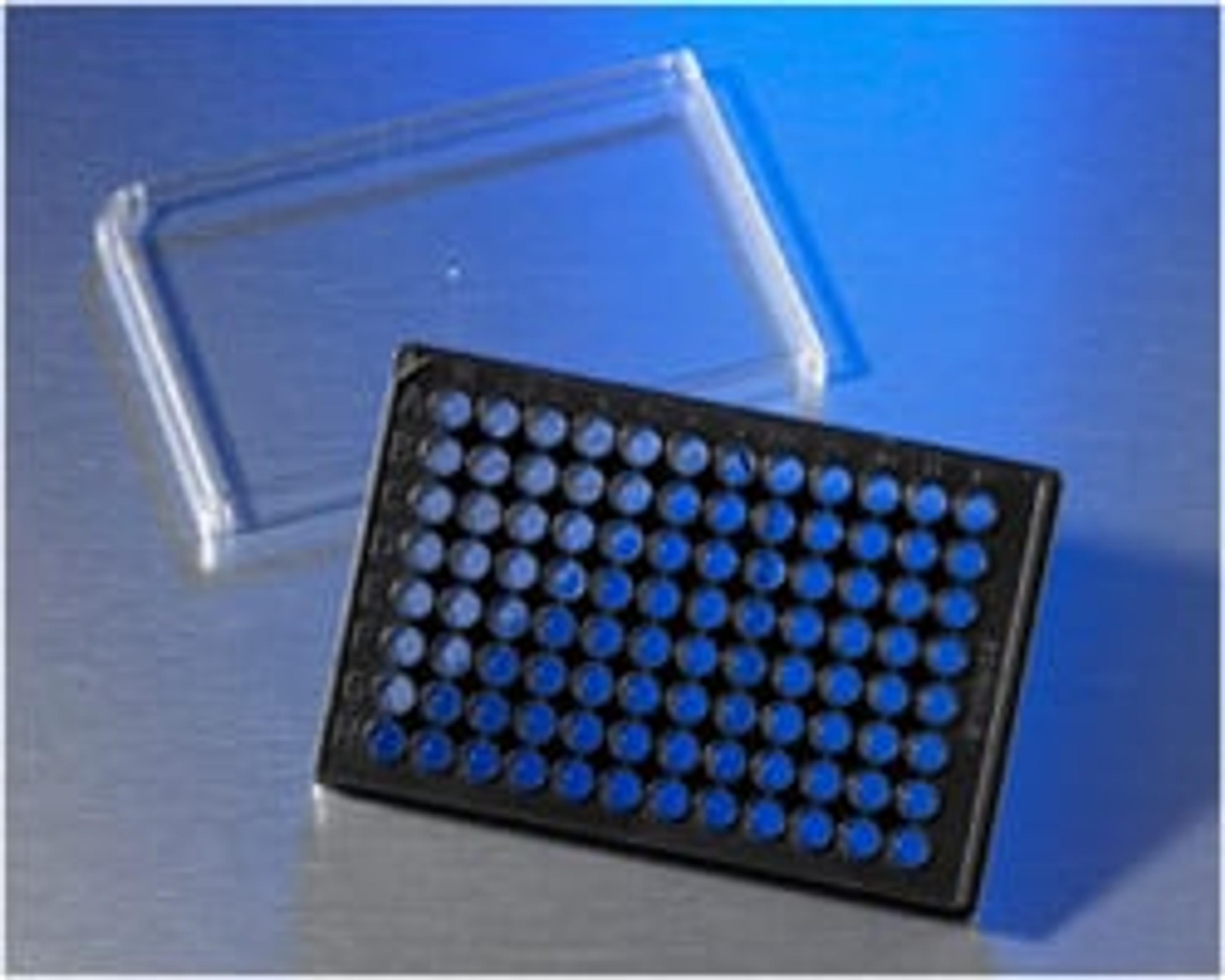
Q: In the spheroid microplate system, are spheroids of equal cell numbers formed?
R.E: Yes, they are formed with equal cell numbers in the spheroid plates, but they can vary with some of the other options available, including magnetic or hanging drop technologies. Careful control of cell number is very important for reproducible well-to-well data, but with the Ultra-Low Attachment plates you can control the cell number fairly easily. As a result of this consistency, you’re able to see a change in the pharmacology that's not reflective of differences in cell number, only to the pharmacology of the compound.
It does become difficult when you grow more than one cell type in a spheroid. And the greater the number of cell types, the greater the differences in cell numbers between spheroids – simply because the cells will grow at different rates. In this context, you need to think about optimizing the media so that it works across numerous cell lines. These kinds of assay are difficult to do, and certainly ones where the assay time will take longer.
Q: What is the optimal size of spheroids for high-throughput screening?
R.E: The cell number in the spheroids will depend upon the plate density that you’re looking at; but let's say you're in a 384-well plate, you would be working with 1,000 to 3,000 cells per spheroid.
The seeding density is irrespective of format and more based on the assay. Smaller spheroids are less likely to be necrotic and are easier to lyse and image with confocal microscopy. Larger spheroids are more likely to have a necrotic core which may be desired. Additionally, larger spheroids result in larger signals for most cell-based assays.
Q: What’s the optimal time that you need to use the spheroids for screening, after cell seeding?
R.E: The optimal time we found with spheroids is roughly between 24 and 48 hours.
Most spheroids form in 24-48 hours but many researchers will culture for much longer. With primary hepatocyte spheroids one of the benefits is that they maintain their functionality for much longer. And then organoids are cultured for several months.
Q: Can you recommend a technique to separate cells in a spheroid into single-cell format to allow for phenotypic analysis with flow cytometry?
R.E: A number of people have tried dropping the calcium levels as well as using a variety of proteases, and titrating the protease to dissociate the cells. Of course, by doing that you lose the spatial location of the cell in this 3D structure and often it's the location of the cell that is very important, either in terms of determining the differentiation state of the cell or the pharmacology of the compound acting on the cell.
That's why I emphasize the importance of imaging, to localize the cell as well as the site of action. The experiment I mention at the beginning of the webinar shows researchers dividing the spheroid into three zones, in order to look at differences in terms of phosphorylation activity between zones within a spheroid.
If the spatial location is not that important, then the lower calcium-type methods and proteases would give you a fairly good dissolution of the cells prior to analysis by flow cytometry.
Q: What technologies do you predict will be game-changing for 3D cell culture in the coming months and years?
R.E: There's a couple on the horizon that are getting everybody excited. One is the growing interest in organoid cell culture. We can now achieve very complicated organoid systems. Some data I've seen suggests that the organoids are truly mimicking the pathology of Alzheimer's disease or Huntington's disease, and the cell architecture is extremely representative. Of course, challenges still exist in this field and organoids must be reproducible well-by-well and week-by-week, but this type of 3D culture is really coming into its own.
We’ve also seen growth of microfluidic technologies to recapitulate the effect of laminar flow in vivo. You can imagine a system where compounds flow from liver cells, through kidney cells, for the analysis of compound metabolism in 3D. There are systems now available where you can even cause the fluid flow to be pulsatile, so it mimics blood flow. The pulsatile activity of fluid flow is known to alter the responsiveness and activity of some cells.
The one technology I suspect that's coming up very quickly is bioprinting, and the ability to use bio-inks to print out 3D tissues with living cells. With this technology, you could print the tissue architecture out exactly how you want it and mimic very complicated tissues, like skin structures for example.