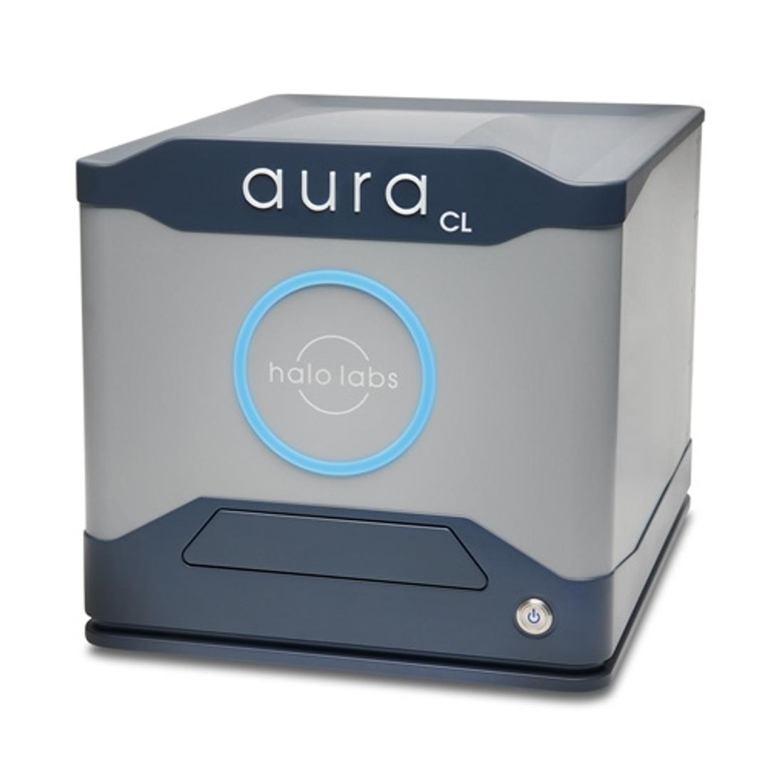
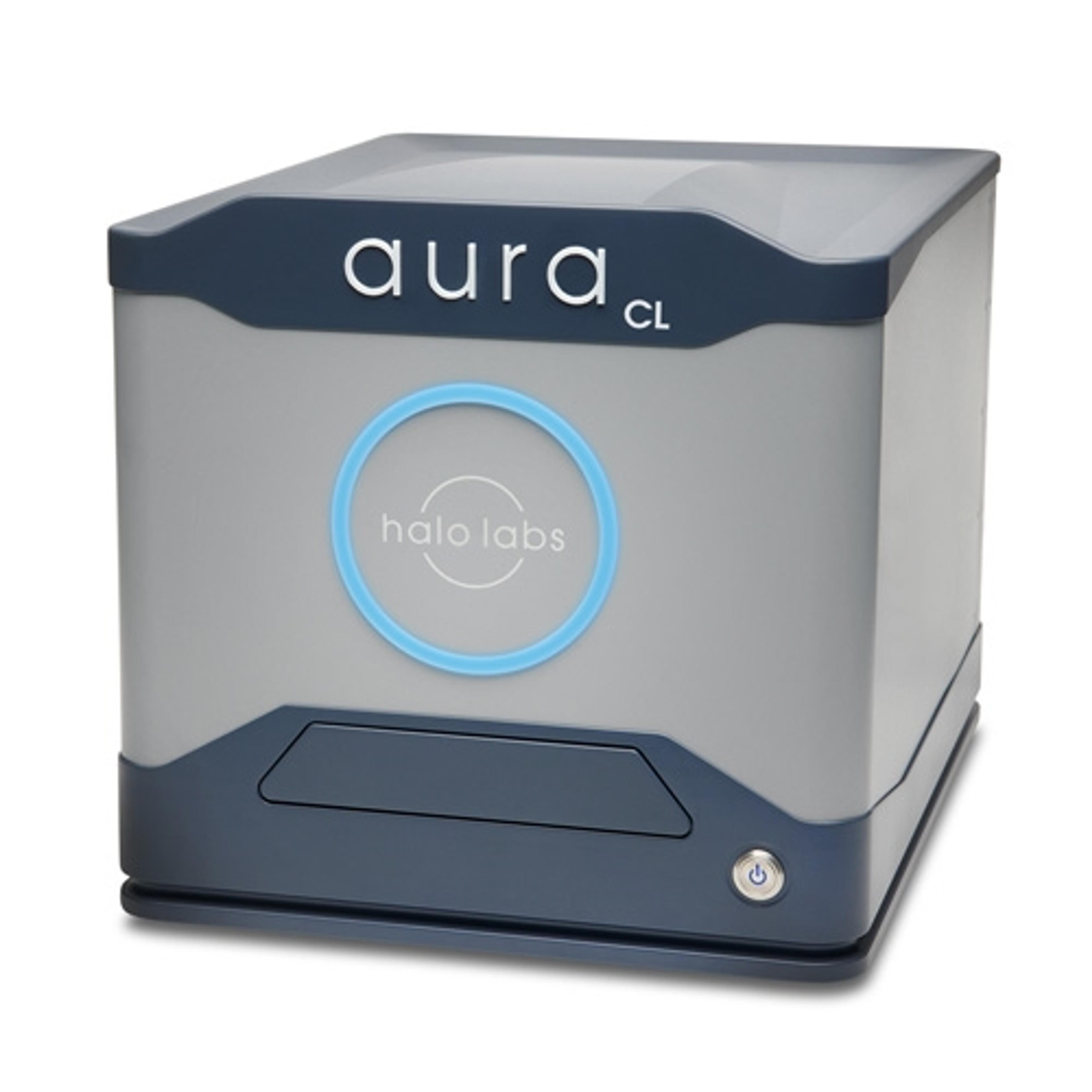
Aura CL
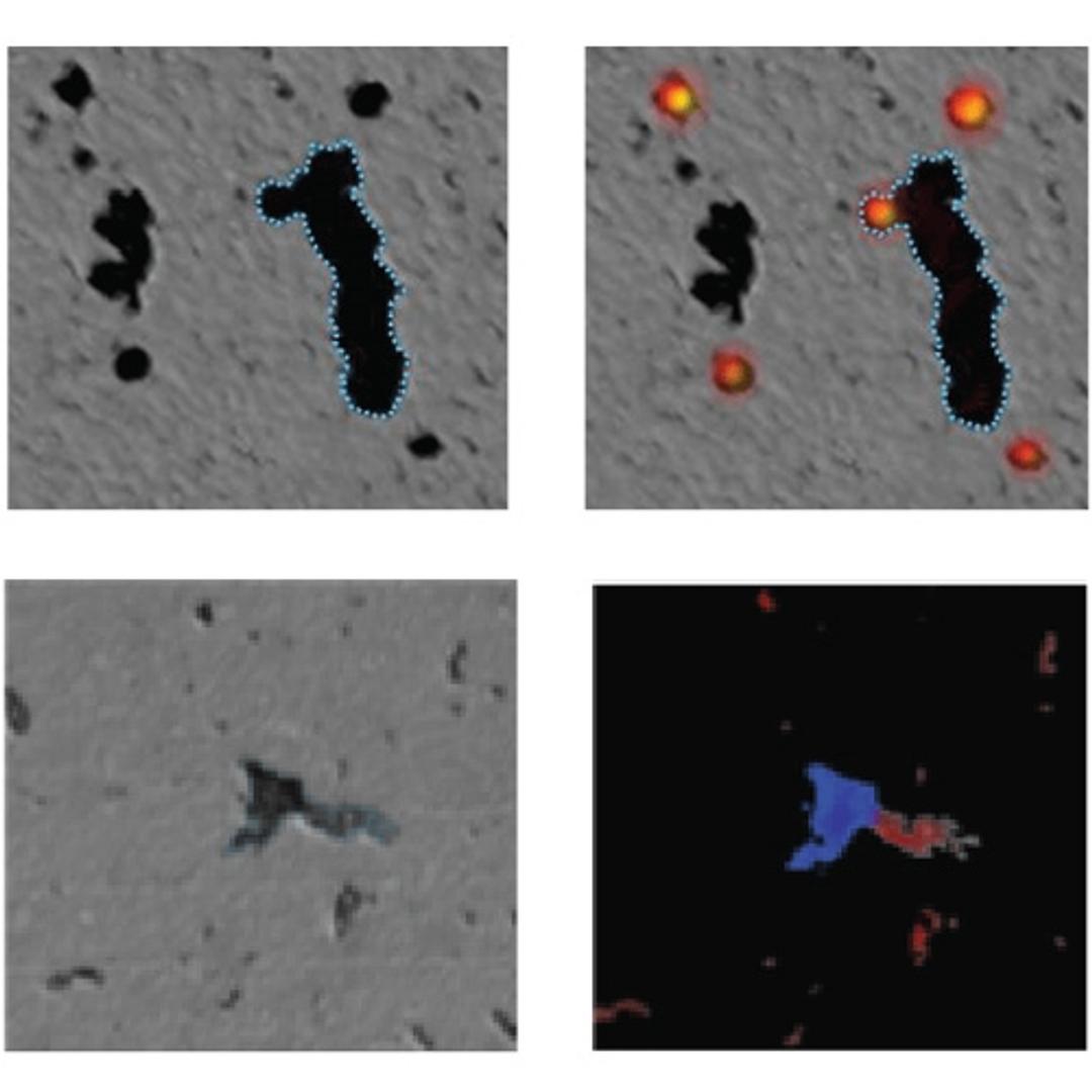
Aura® CL systems couples Fluorescent Membrane Microscopy and Backgrounded Membrane Imaging to help you understand what’s in your cell therapy. It detects, counts, sizes, and IDs cellular aggregates and subvisible particles and ID’s cellular from other particles without any machine learning so you can maximize the purity, safety, and efficacy of your therapeutic.

The supplier does not provide quotations for this product through SelectScience. You can search for similar products in our Product Directory.
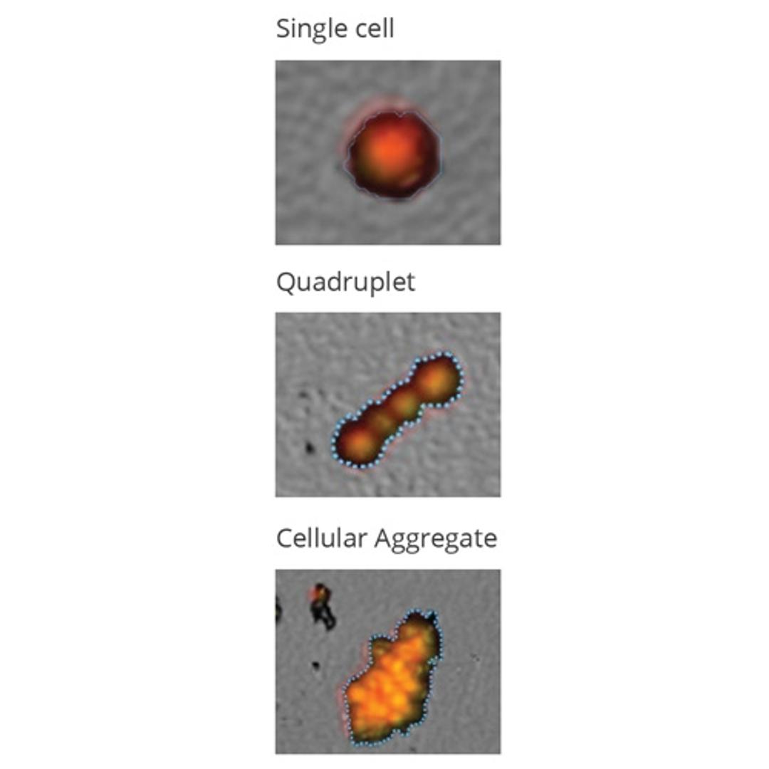
Aura® CL is the first system designed to detect, count, and characterize cellular aggregates and subvisible particles for product quality measurements in cell therapy applications. It also makes it super simple for you to specifically ID cell from non-cell aggregates right out of the box, without having to spend hours sorting through images or needing complex machine learning libraries.
Aura CL combines Backgrounded Membrane Imaging (BMI) with two channels of Fluorescence Membrane Microscopy (FMM) to give you aggregate data without any clogging concerns or the need to clean between measurements. Get count, size, and morphological information using BMI with full-well imaging and 100% sampling efficiency, or differentiate between cellular, protein, or extrinsic aggregates using FMM to quickly know what’s in your sample. FMM can also be used for cell viability and cell type differentiation assays to further understand your cell therapy product.
- Cell aggregate identification and differentiation
- Identify, quantitate, and differentiate between proteinaceous and non-proteinaceous materials
- Only requires 5uL of sample volume
- Fluorescence labeling of your choice
- Completely automated and fluidic free with minimal optimization
- Large Size Particle Detection Range: 1µm – 5mm 96 sample throughput
- Established USP method

