CellPathfinder
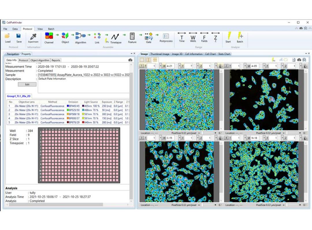
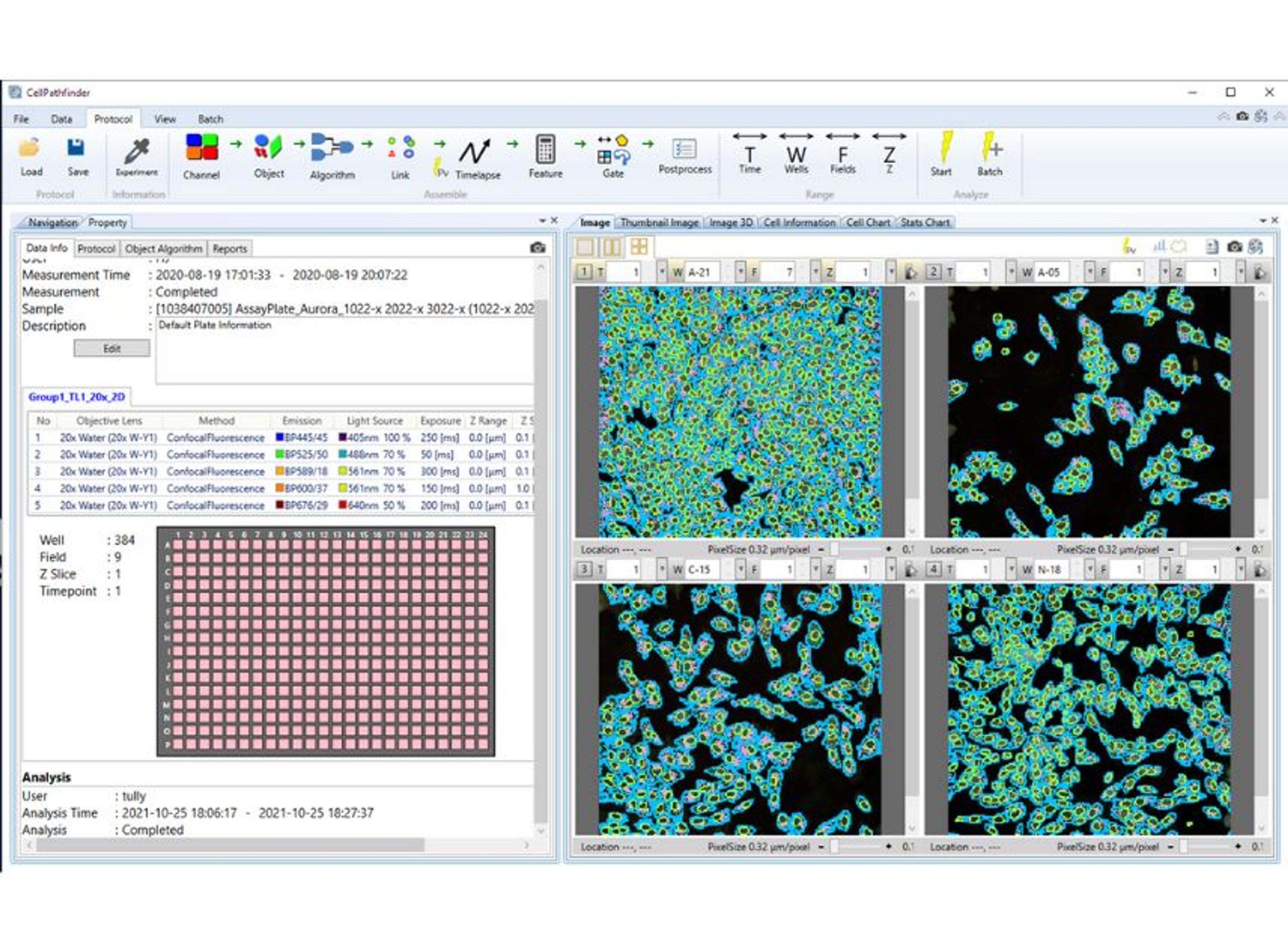
Easy to use software for automated primary image analysis, phenotyping, live cell tracking and cellular assays. Intuitive and powerful high content analysis software for primary image analysis and graphing for lead optimization and phenotyping with deep learning tools. Provides users with advanced insights to complex analysis questions.

The supplier does not provide quotations for this product through SelectScience. You can search for similar products in our Product Directory.
Great software for a high use, multidisciplinary core facility
High-content screening
CellPathfinder is a wonderful analysis software. Last year we did a head to head among all high-content instruments on the market and decided on the CQ1 in part because our users found the software to hit that sweet spot of incredibly powerful yet easy to use. It was also the only software that allowed us to export our data as .FCS files so they could be further processed in our existing bioinformatics pipelines. As an added bonus, I was also able to teach my 5 year old son how to use the software so he can take "cool" pictures of the bugs he finds.
Review Date: 29 Aug 2024 | Yokogawa Corp. of America
Good and easy to use product
Segmentation of cell microscopy fluorescence images
I loved how easy it was to generate segmentation for a biologist like me who didn't have formal coding training. The pipelines I created were very similar in accuracy to what our dedicated computational scientist would output and that made me feel confident I could analyze my own data internally for next steps.
Review Date: 29 Aug 2024 | Yokogawa Corp. of America
Yokogawa’s CellPathfinder Analysis Software is a powerful tool designed to revolutionize how you interpret and analyze complex cellular data. Tailored for high-content screening and high-resolution imaging applications, CellPathfinder offers an extensive suite of features to provide insights into cellular processes, morphology, and phenotypic variations. At the heart of CellPathfinder is its advanced image analysis engine, which delivers precise and reproducible results. The software excels in automated cell segmentation and classification, enabling researchers to quickly and accurately identify and quantify cells and subcellular structures across large datasets.
CellPathfinder user-friendly interface is designed for both novice and experienced users, providing intuitive navigation and streamlined workflows. The software supports a wide range of imaging modalities, including fluorescence, brightfield, and phase contrast, making it versatile and adaptable to various research needs. Its flexible analysis tools can be customized to meet specific experimental requirements, offering a high degree of adaptability in complex studies.
CellPathfinder also integrates seamlessly with Yokogawa’s high content imaging systems, ensuring a smooth workflow from image acquisition to data analysis. The software’s robust data management features support large-scale studies by providing efficient storage, retrieval, and visualization of complex datasets. Researchers can easily access and compare results across different experimental conditions, facilitating more thorough and informed analysis.
The software’s advanced statistical and graphical tools further enhance its analytical capabilities. With built-in options for statistical analysis, heat maps, scatter plots, and other visual representations, CellPathfinder helps researchers interpret complex data and identify trends with clarity. These tools are essential for validating findings, generating publication-quality figures, and communicating results effectively.
Overall, Yokogawa's CellPathfinder software is a powerful and user-friendly tool that empowers researchers to efficiently analyze complex cellular data and gain valuable insights into their biological processes.
Key Features and Benefits:
- Intuitive interface guides users through processes
- Provides abundant analysis functions to customize segmentation
- Supports label-free segmentation
- Multiple graphing tools with a streamlined, easy-to-use interface
- Easy image comparison improves efficiency and reduces errors
- Advanced analysis with Deep Learning Module
- Easy and flexible graph generation
- Yokogawa’s proprietary image generation technology “CE Bright Field” leads to label-free analysis of samples
- Easy-to-use machine-learning
Assay and Applications:
- Cell counting and colony counting
- 3D analysis of spheroids and/or organoids
- Lipid droplets
- Apoptosis
- Neurite outgrowth
- Migration and cell tracking
- FISH
- Cell Painting
- Cell cycle and spindle studies
- Phase contrast analysis
- Autophagy
- Intracellular organelle localization
- Calcium flux and other fast timelapse analysis
- Live/Dead
- Nuclear Translocation
- Angiogenesis
Not available in the following countries: North Korea, Iran, Iraq, Libya, Cuba, Syria, Sudan, South Sudan, Afghanistan, Democratic Republic of Congo, Central African Republic, Somalia, Lebanon, India, Pakistan, Russian Federation, Republic of Belarus, Ukraine.

