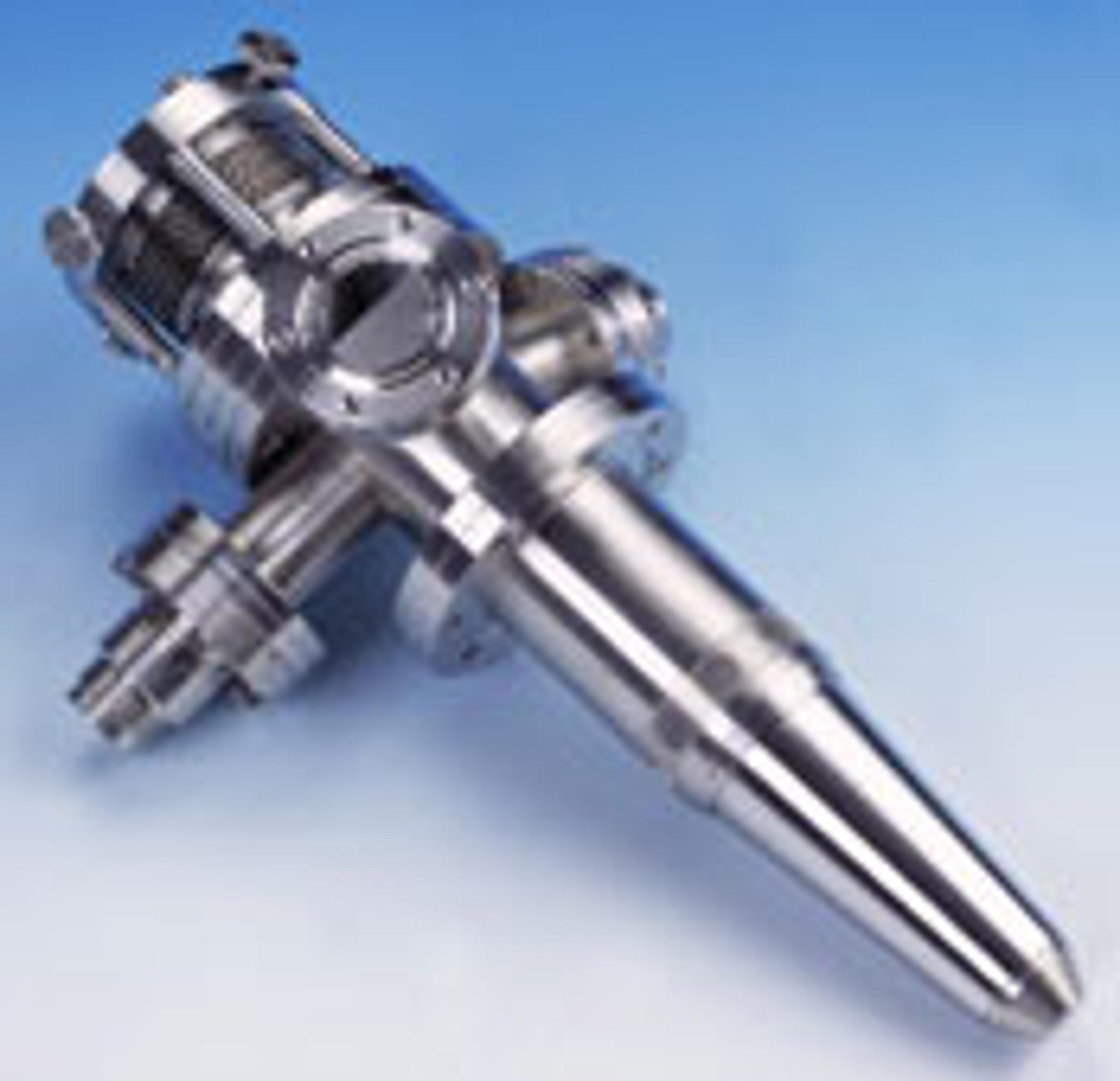
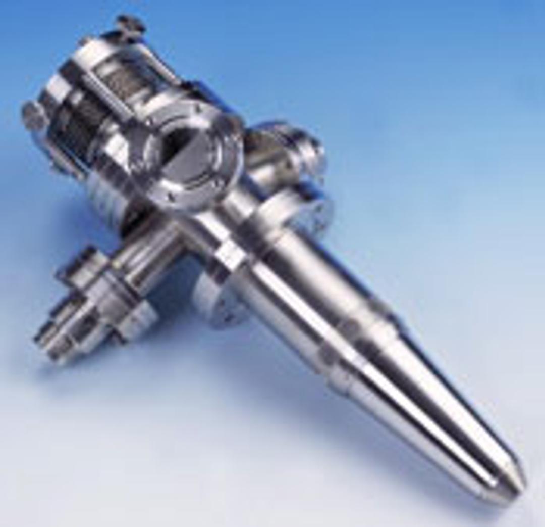
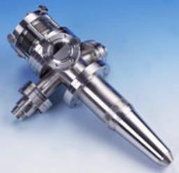
FEG 1000 Field Emission Source Electron Gun
The FEG1000 is optimised for Auger analysis at very high spatial resolution with SEM and SAM imaging: High brightness Schotty field emission source High current density 100 nm spot size at 5 nA sample current Fully integrated with the Avantage Windows NT based data system for operation and scanning Product detail: High Resolution, Field Emission Source Electron Gun for SEM, AES and SAM High brightness Schotty field emission…

The supplier does not provide quotations for this product through SelectScience. You can search for similar products in our Product Directory.
The FEG1000 is optimised for Auger analysis at very high spatial resolution with SEM and SAM imaging:
High brightness Schotty field emission source
High current density
100 nm spot size at 5 nA sample current
Fully integrated with the Avantage Windows NT based data system for operation and scanning
Product detail:
High Resolution, Field Emission Source Electron Gun for SEM, AES and SAM
High brightness Schotty field emission source
High current density
100 nm spot size at 5 nA sample current
Fully integrated with the Avantage Windows NT based data system for operation and scanning
Magnetically shielded construction
The FEG1000 features a field emission source for maximum current density and produces a 100 nm spot size at 5 nA. Optimised for Auger analysis at very high spatial resolution with SEM and SAM imaging, the gun is available with a PC-based data system.
Source
The electron source is a Schotty field emitter (ZrO/W). This type of source delivers a bright, highly-stable electron beam into the optical column. To ensure the stable operation of the field emission source, the gun must be differentially pumped. An ion pump is supplied as part of the FEG1000 package.
Optical Column
The FEG1000 has a two-lens column, a condenser lens and a final focus lens. These allow the user to select the current/spot size combination required for the analysis.
Beam Forming
The electron beam is focused onto the sample using a single electrostatic lens. The beam quality is optimised using the octopole stigmator.
Scanning
A quadrupole arrangement of electrodes is used to scan the electron beam. This compact arrangement minimises the distance from the focusing lens to the sample so that the smallest spot size can be achieved.
Magnetic Shielding
Mu-metal shielding is fitted in sensitive areas to prevent stray magnetic fields from affecting the beam quality or direction.