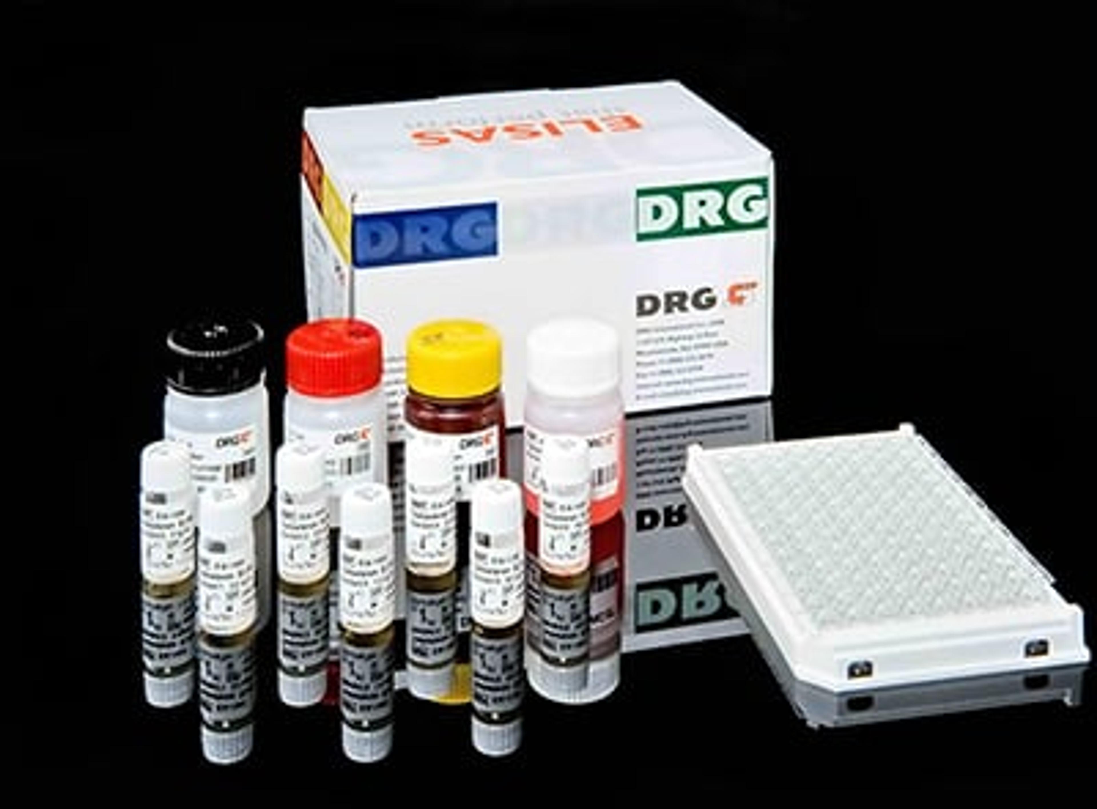
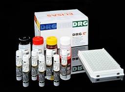
TGF-B2 ELISA
High Quality Assays with Reproducible and Reliable Results


The supplier does not provide quotations for this product through SelectScience. You can search for similar products in our Product Directory.
An enzyme immunoassay for the quantitative measurement of transforming growth factor Beta2 (TGF-Beta2) in serum, plasma, and cell culture supernatant. Transforming growth factor-Beta (TGF-Beta) is a multipotent Cytokine with cell- and dose-dependent activities. This molecule is produced by a number of cells and tissue types, e.g.thrombocytes, bone tissue, placenta and kidneys. TGF-Beta2 is not produced by blood platelets, contrary to TGF-Beta1. This potent Cytokine modulates embryonic development, bone formation, mammary development, wound healing, hematopoiesis, cell cycle progression and the production of the extracellcular matrix. TGF-Beta2– null mice were shown to exhibit perinatal mortality and a wide range of developmental defects for a single gene description which include cardiac, lung, craniofacial, limb, spinal column, eye, inner ear and urogenitial defects. TGF-Beta2 has been shown to be a potent growth inhibit factor in the regulation of postnatal cerebellar neurons and neuroblast proliferation. TGF-Beta2 has beendetected in tear fluid. TGF-Beta2 levels are elevated in the vitreous of patients with proliferative diabetic retinopathy. Elevated plasma levels of TGF-Beta2 have been described in patients with disseminated malignant melanoma. TGF-Beta2 concentrations are furthermore elevated in Parkinson’s disease in ventricular cerebrospinal fluid. The DRG TGF-Beta2 ELISA Kit is a solid phase enzyme-linked immunosorbent assay (ELISA) based on the sandwich principle. Prior to testing the standards and patient samples are diluted in assay buffer, acidified with HCl and then neutralized with NaOH. Afterwards, the neutralized standards and samples are added to the antibody coated (polyclonal) microtiter wells. After the first incubation the unbound sample material is removed by washing. Then a biotinilated mouse anti TGF-Beta2 antibody and the Streptavidin-HRP Enzyme complex are incubated in succession. An immuno enzyme sandwich complex is formed. After incubation the unbound conjugate is washed off. Having added the substrate solution, the intensity of colour developed is proportional to the concentration of TGF-Beta2 in the patient sample.