THUNDER Imager Tissue
Real-time fluorescence imaging of 3D tissue sections typically used in neuroscience and histology research
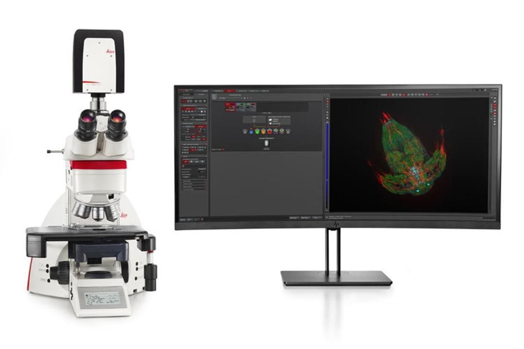
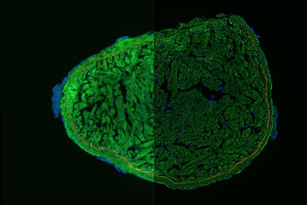
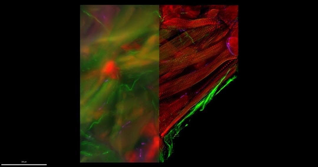
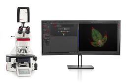
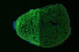
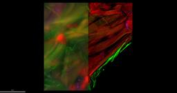
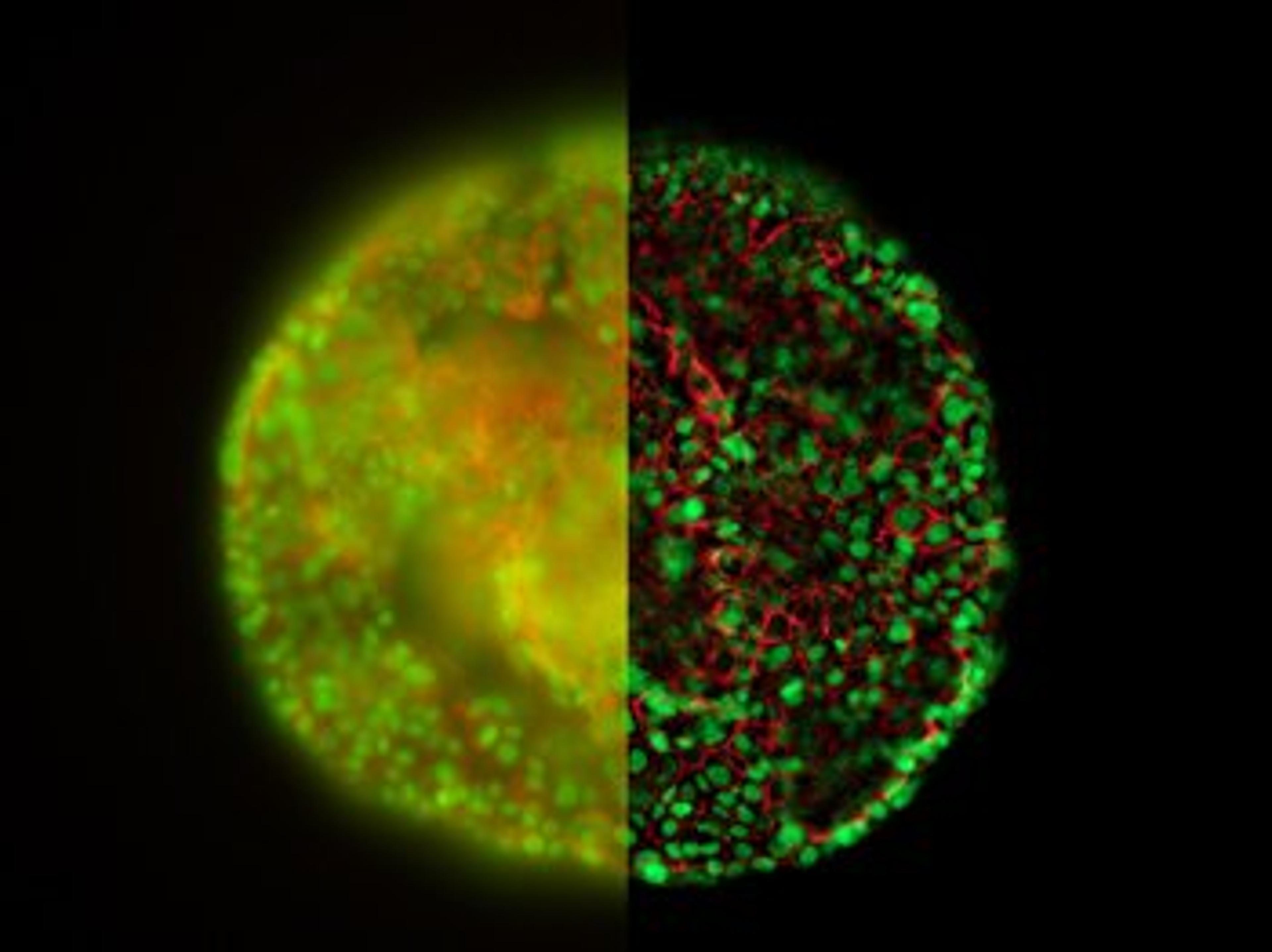
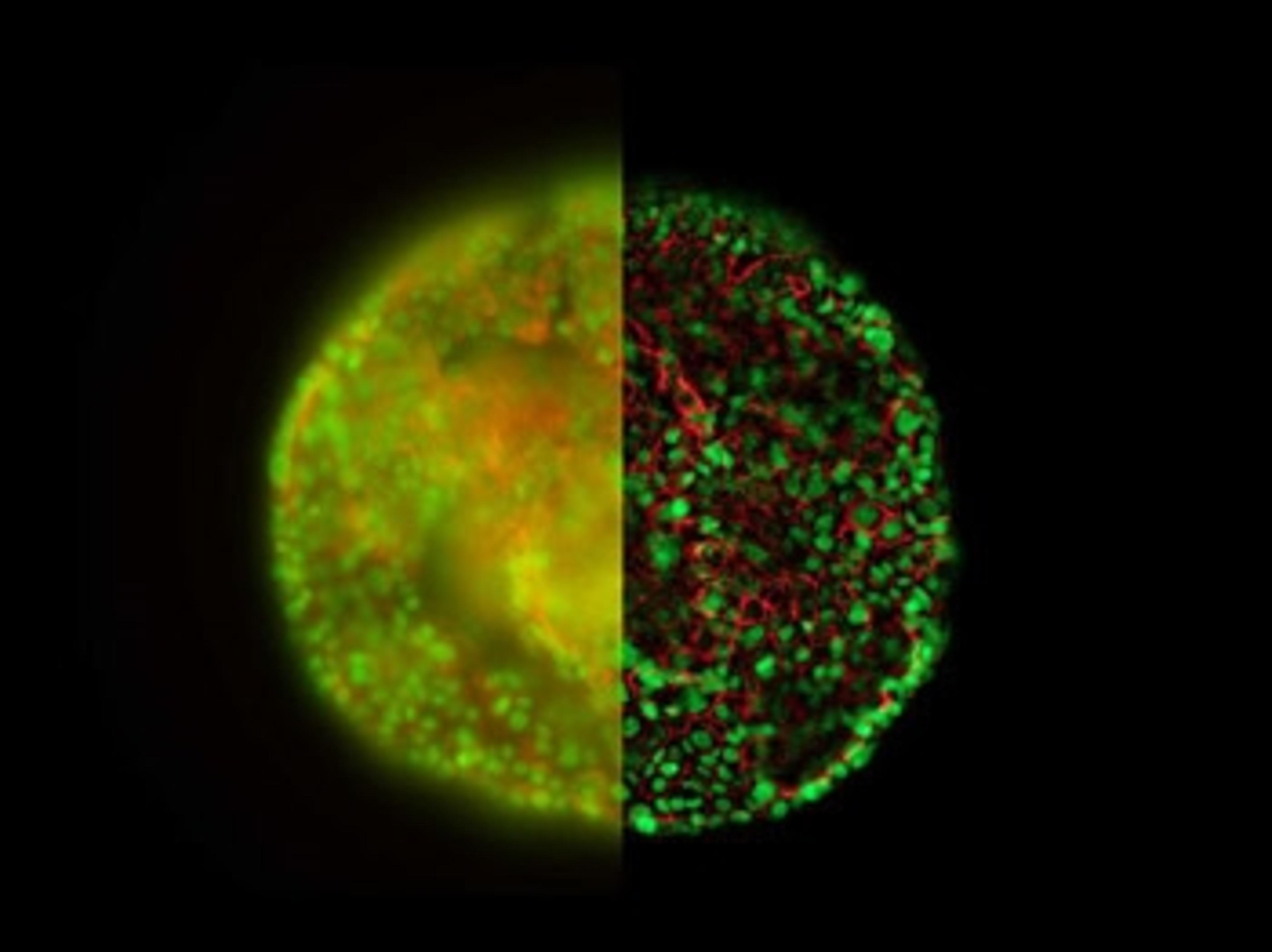
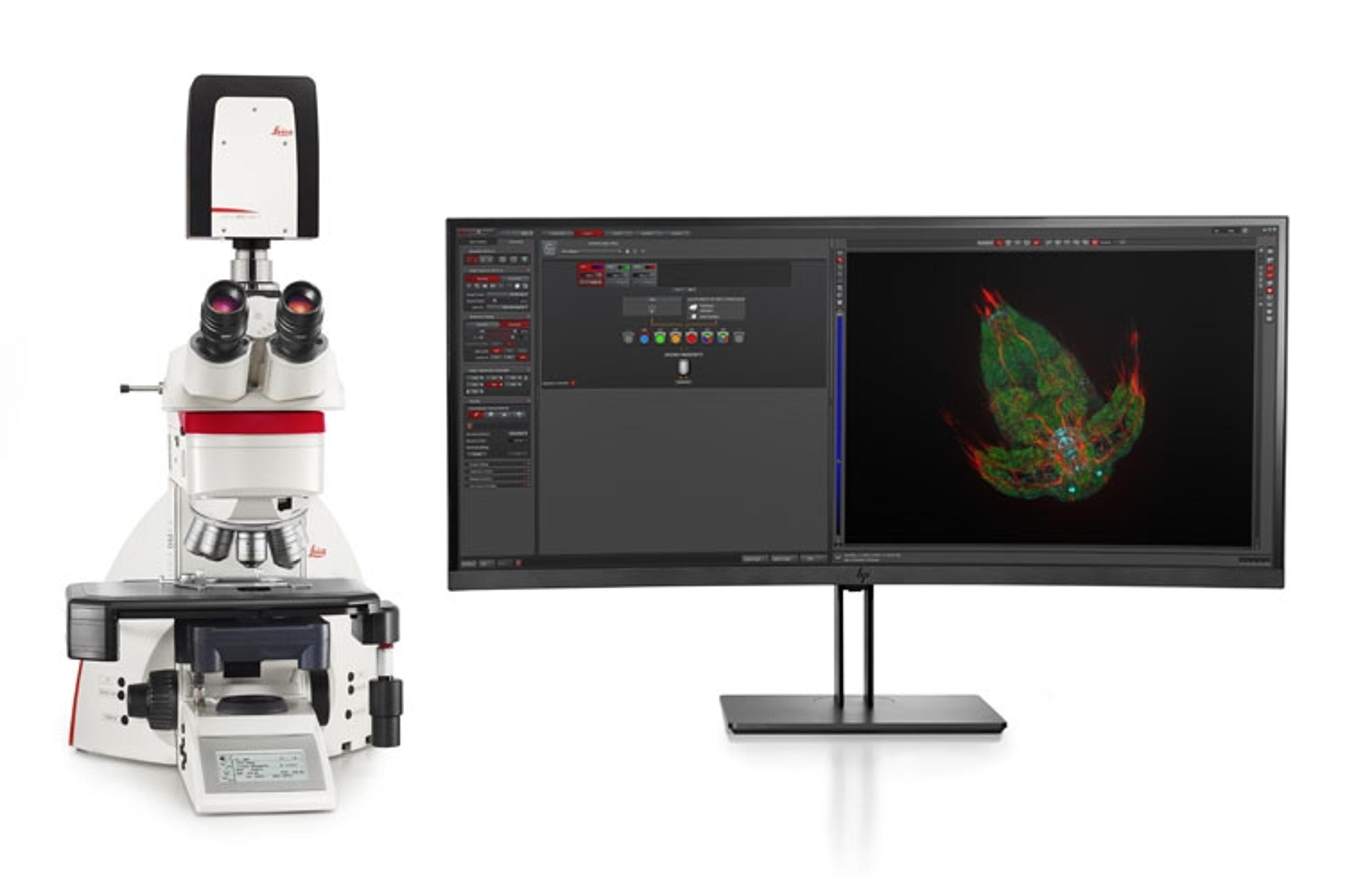
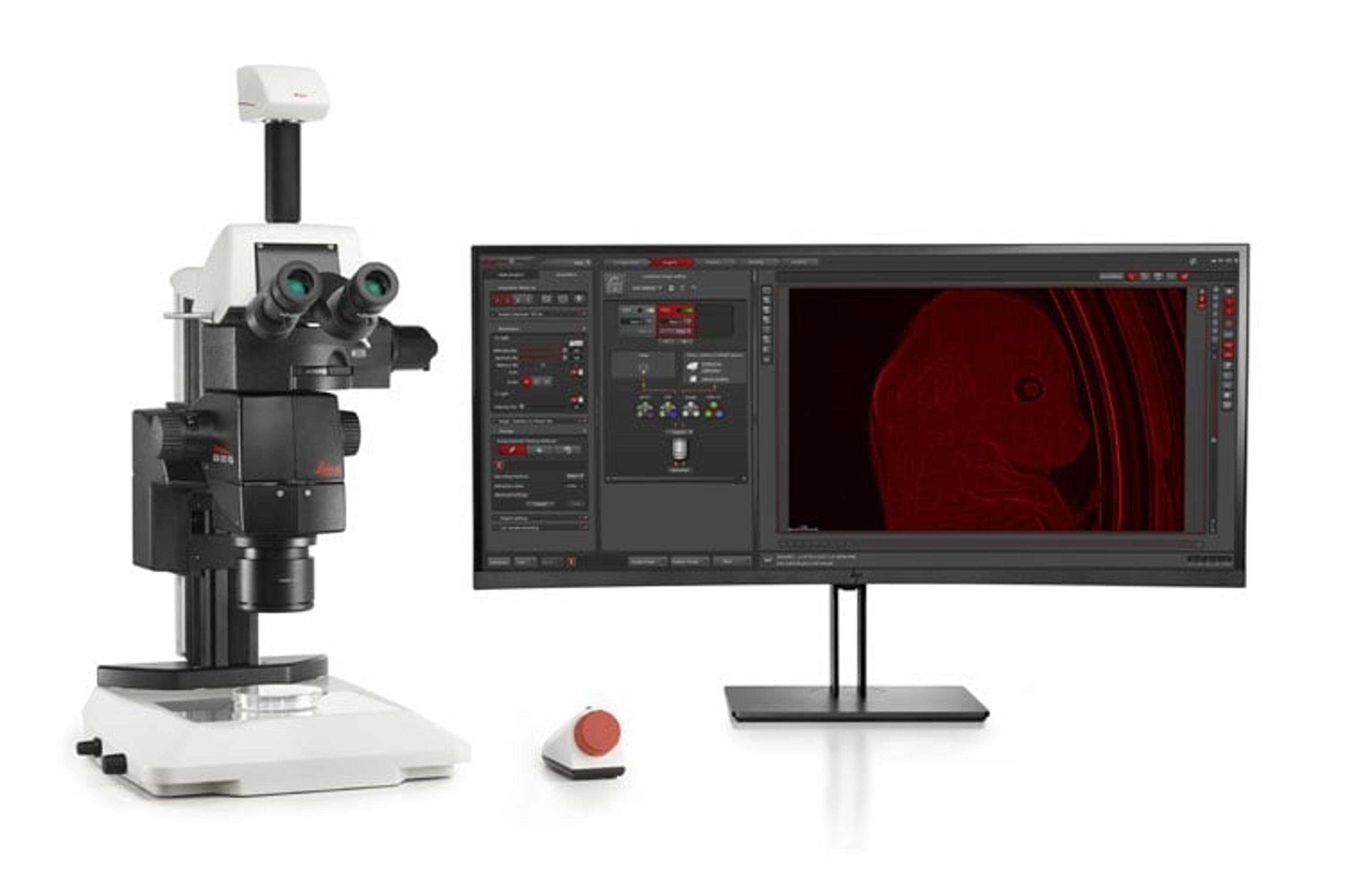
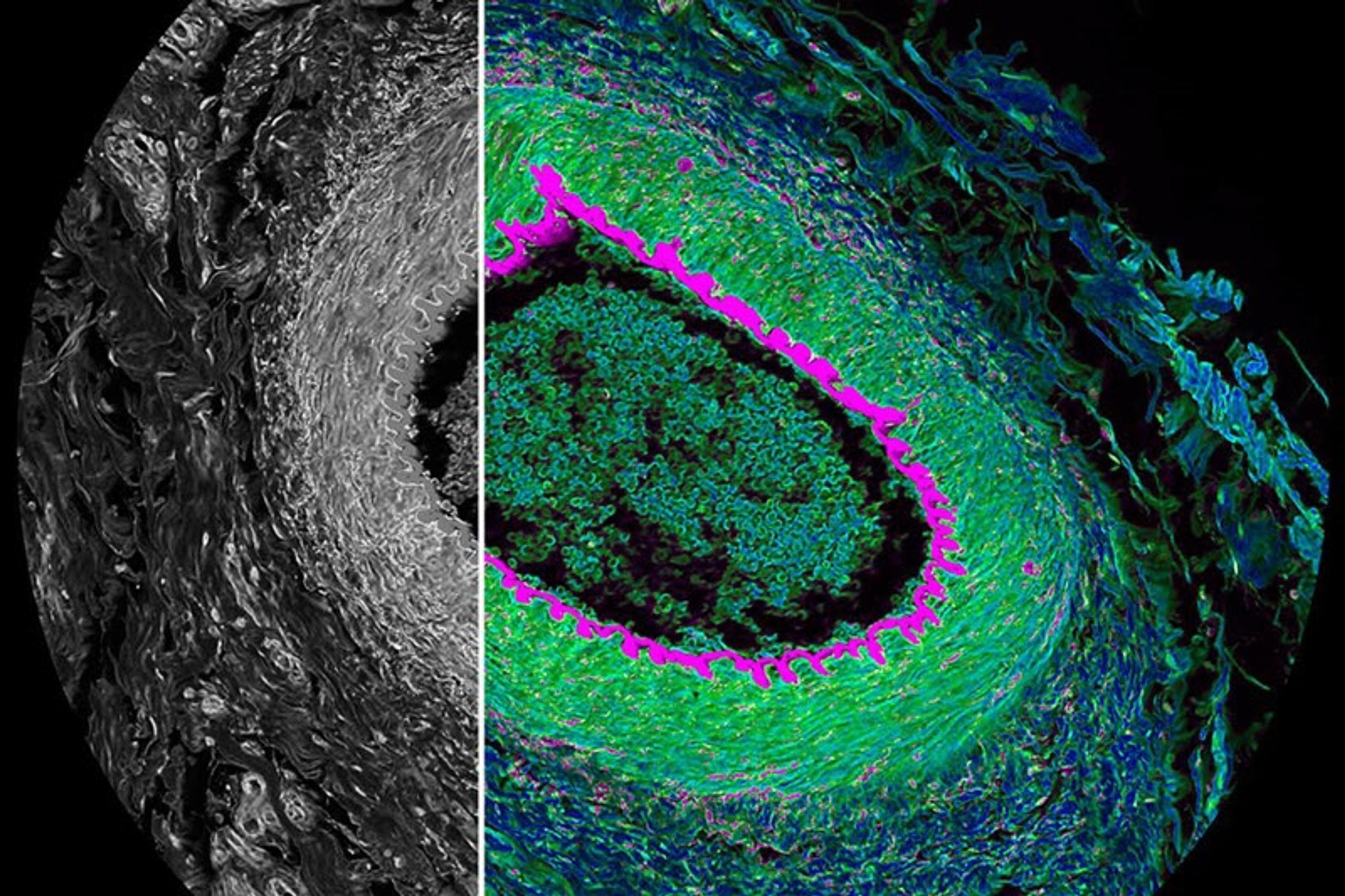
The THUNDER Imager Tissue allows real-time fluorescence imaging of 3D tissue sections typically used in neuroscience and histology research. Acquire rich, detailed images of thick tissues free of haze from out-of-focus blur. Even fine structures deep in tissues can be resolved thanks to Computational Clearing, an innovative Leica technology. Image detailed morphological structures like axons and dendrites of neurons in a brain slice. The high image quality, even with thick tissue sections, is combined with the well-known speed, fluorescence efficiency, and ease of use of widefield microscopes.
Advantages for your research are:
- Rapidly acquire blur-free images showing finest details of the morphology, even deep within thick sections
- Get fast overviews of whole tissue sections
- Image and analyze challenging tissue sections with an easy workflow
THUNDER Imagers feature the innovative Leica technology Computational Clearing. It efficiently removes out-of-focus blur in real time, enabling the meaningful use of 3D specimens with camera-based fluorescence microscopes. The high sensitivity of the system ensures low phototoxicity and photobleaching, i.e., higher throughput with optimal conditions.
Find the THUNDER Imager that’s right for you
The THUNDER Imager Tissue is part of the THUNDER family of imaging systems. Whether you are looking for a dedicated high-end imaging system that excels in a given application, or a versatile solution for a lab running different kinds of assays with various samples, we’ve got you covered.

