The nVista System for Neural Circuit Imaging
nVista is the first end-to-end solution for visualizing large-scale neural circuit dynamics in freely behaving animals.

The supplier does not provide quotations for this product through SelectScience. You can search for similar products in our Product Directory.
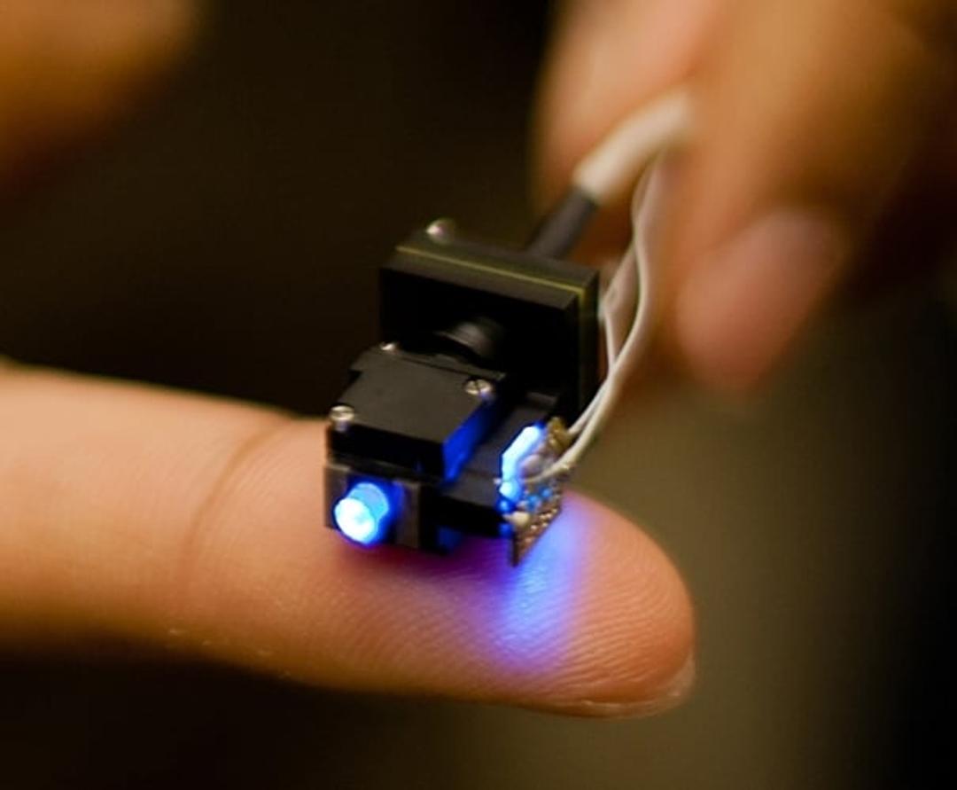
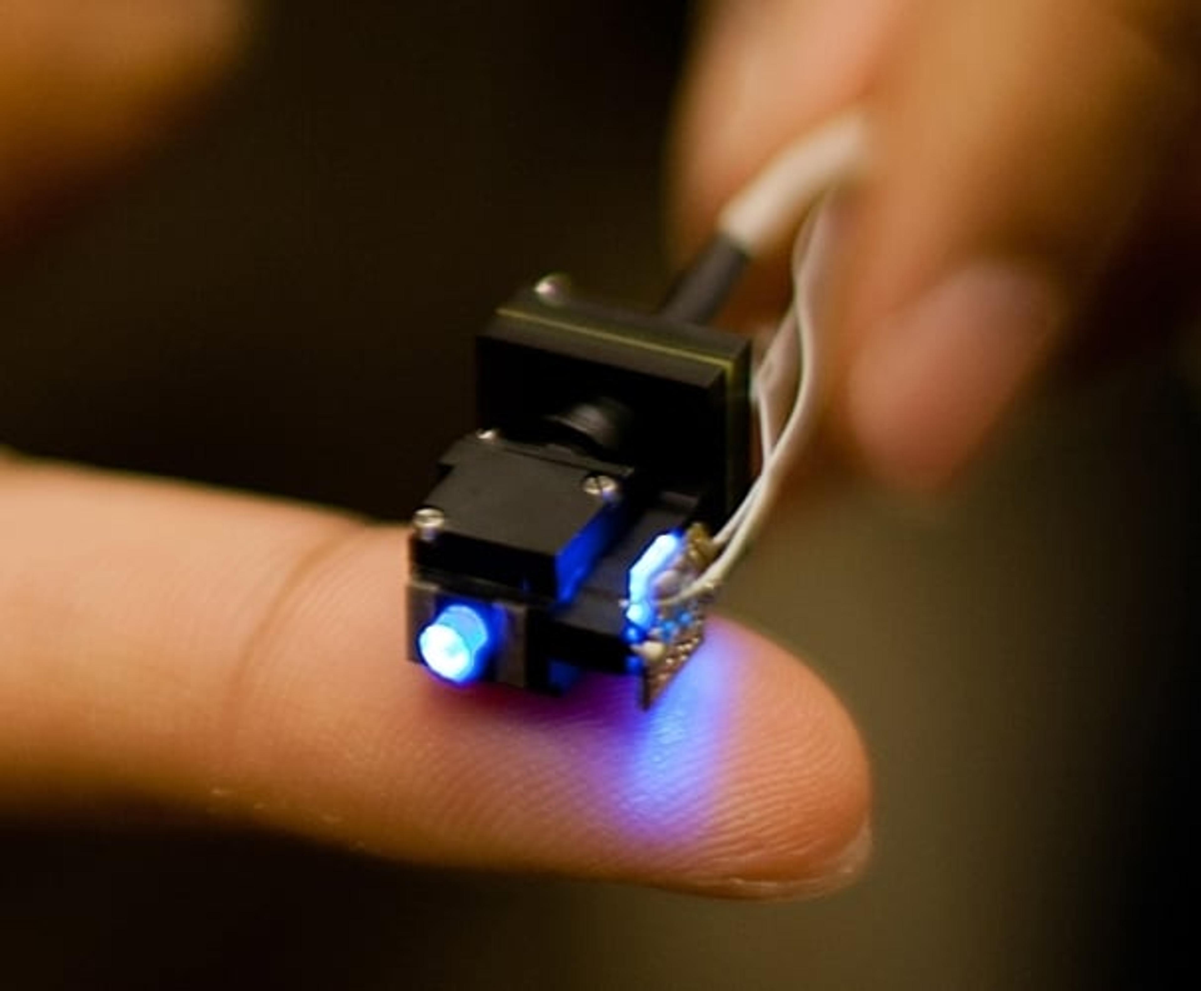
Introducing the world's smallest neural circuit imaging system designed to obtain Ca2+ imaging data from hundreds of neurons in awake, behaving animals in almost any brain region you can imagine. Proven through high impact journal publications and used in top labs around the world, nVista’s core miniature microscope technology lets you uncover new neural circuit insights in the active brain.
A complete solution for the neural circuit researcher, the nVista System comprises our 2 gram head-mounted miniaturized microscope; data acquisition hardware to sync and trigger with external devices; the nVista Data Acquisition Software to record your experimental data; and our data analysis platform Mosaic, whose intuitive apps streamline the processing of your raw imaging data. The nVista System is backed by a world-class Support team, offering unparalleled technical and scientific guidance and training to help you overcome barriers to success in the lab.
Whether you’re an experienced electrophysiologist or a savvy brain imaging expert, nVista will complement your research program. nVista enables:
- LARGE FIELD OF VIEW WITH SINGLE-CELL RESOLUTION:
Visualize and record population-scale neural activity with cellular resolution in pre-determined cell types. - ROBUST LONGITUDINAL STUDIES:
Perform chronic imaging studies in freely behaving subjects. - NATURAL ANIMAL BEHAVIOR:
Plug into standard behavioral assays to perform time-locked imaging and behavioral experiments. - EXPERIMENTAL FLEXIBILITY:
Image any brain area, including deep brain structures and surface brain regions. Use the latest, fastest genetically-encoded Ca2+ indicators.
Microscope Specifications:
Mass 2 g Dimensions 11 mm x 14 mm x 20 mm Modality Single-channel epifluorescence Excitation 475/10 nm (blue) Collection 535/50 nm (green) Cable Length 2.5 m Maximum Field of View 1440 pixels x 1080 pixels; 900 μm x 650 μm Temporal Resolution 30 fps at full field of view with USB 3.0
Recent Publications:
Kitamura et al. Entorhinal Cortical Ocean Cells Encode Specific Contexts and Drive Context-Specific Fear Memory. Neuron (2015)
Sun, et al. Distinct speed dependence of entorhinal island and ocean cells, including respective grid cells. PNAS (2015)
Pinto, et al. Cell-Type-Specific Activity in Prefrontal Cortex during Goal-Directed Behavior. Neuron (2015)
Markowitz, et al. Mesoscopic Patterns of Neural Activity Support Songbird Cortical Sequences. Plos Biology (2015)
Betley, et al. Neurons for hunger and thirst transmit a negative-valence teaching signal. Nature (2015)
Jennings, at al. Visualizing Hypothalamic Network Dynamics for Appetitive and Consummatory Behaviors. Cell (2015)

