Light Microscopy Products & Reviews

25

Light microscopes or optical microscopes are used to visualize microscale objects under magnification, including cells, clinical specimens and materials. Lab equipment for light microscopy includes confocal microscopes, fluorescence microscopes, zoom and stereo microscopes. Microscope slides and imaging reagents are available for visualizing samples, as well as various microscope stages and incubators for large or temperature-sensitive samples. Find the best light microscopes in our peer-reviewed product directory: compare products, check customer reviews and receive pricing direct from manufacturers.

Product Filter

Explore by Field
Light Microscopy
Refine your selection
Explore by Company

Ratings

Search

Selected Filters:

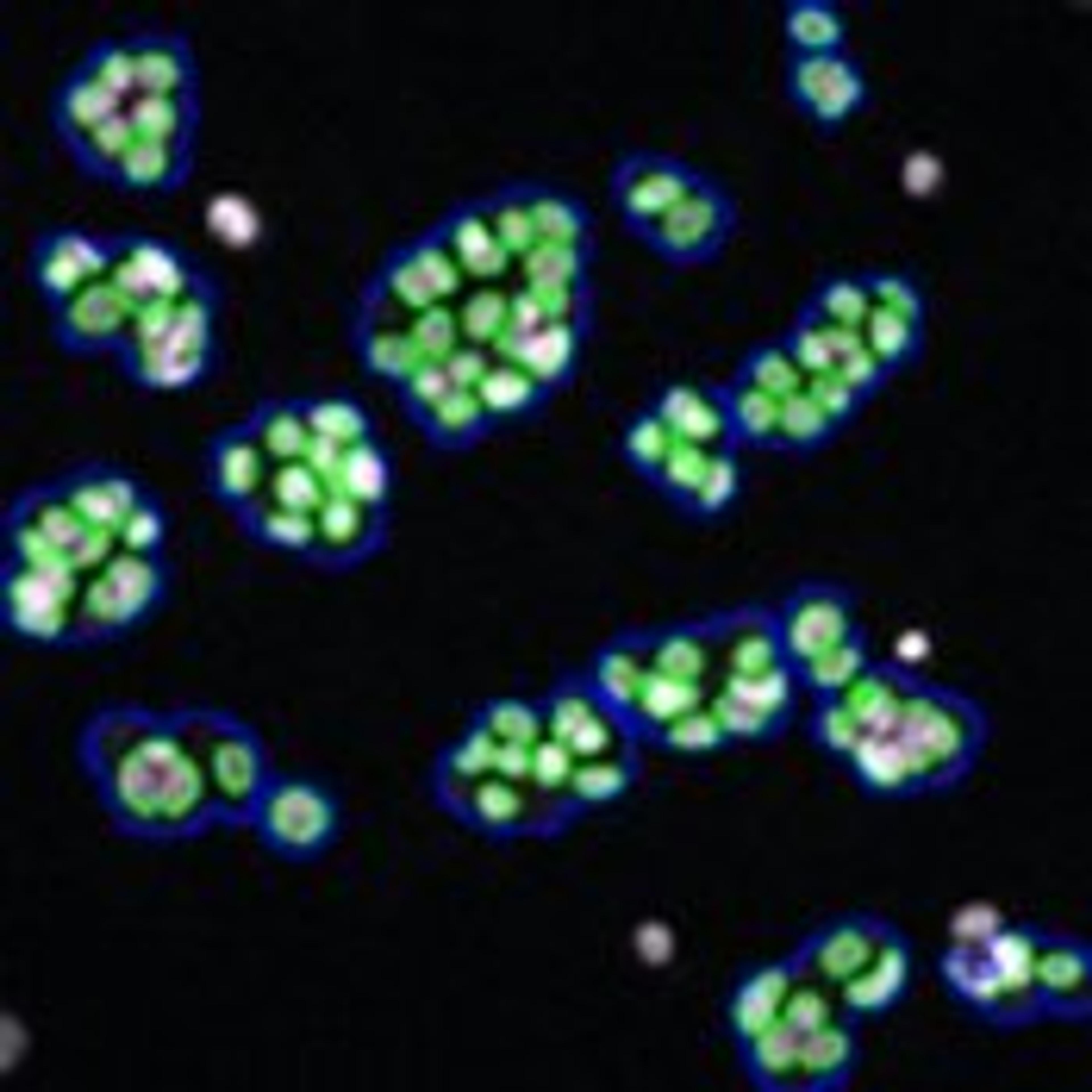
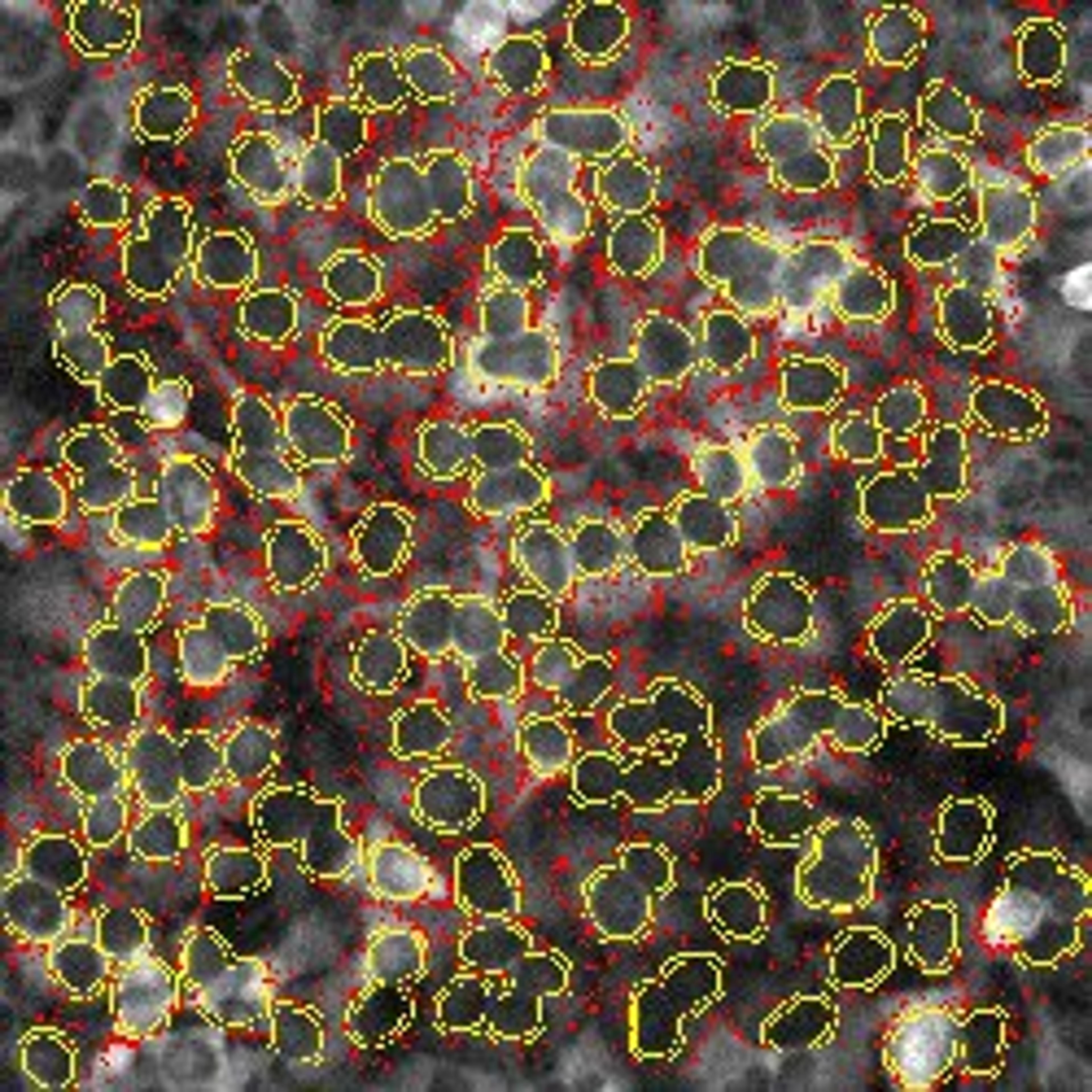
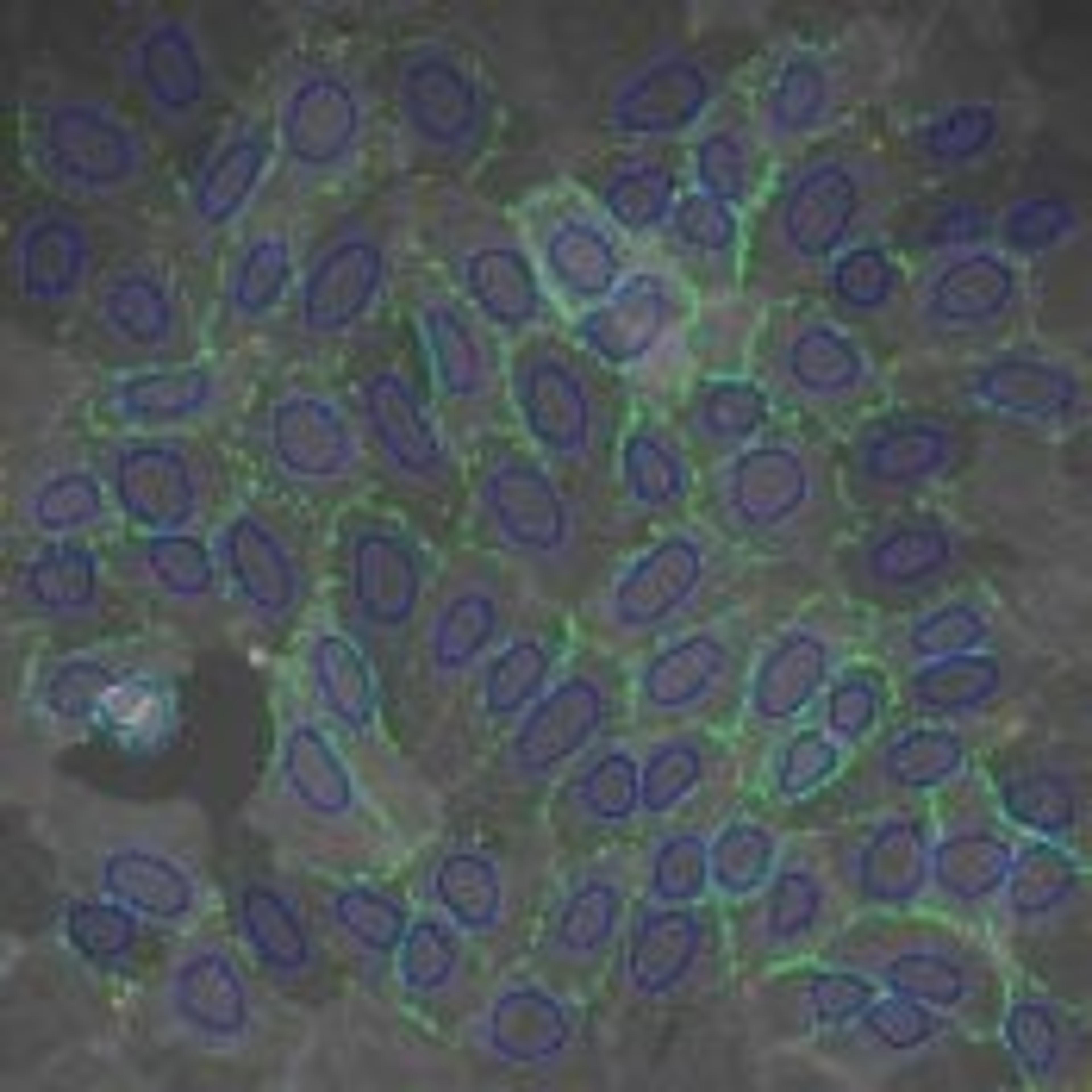
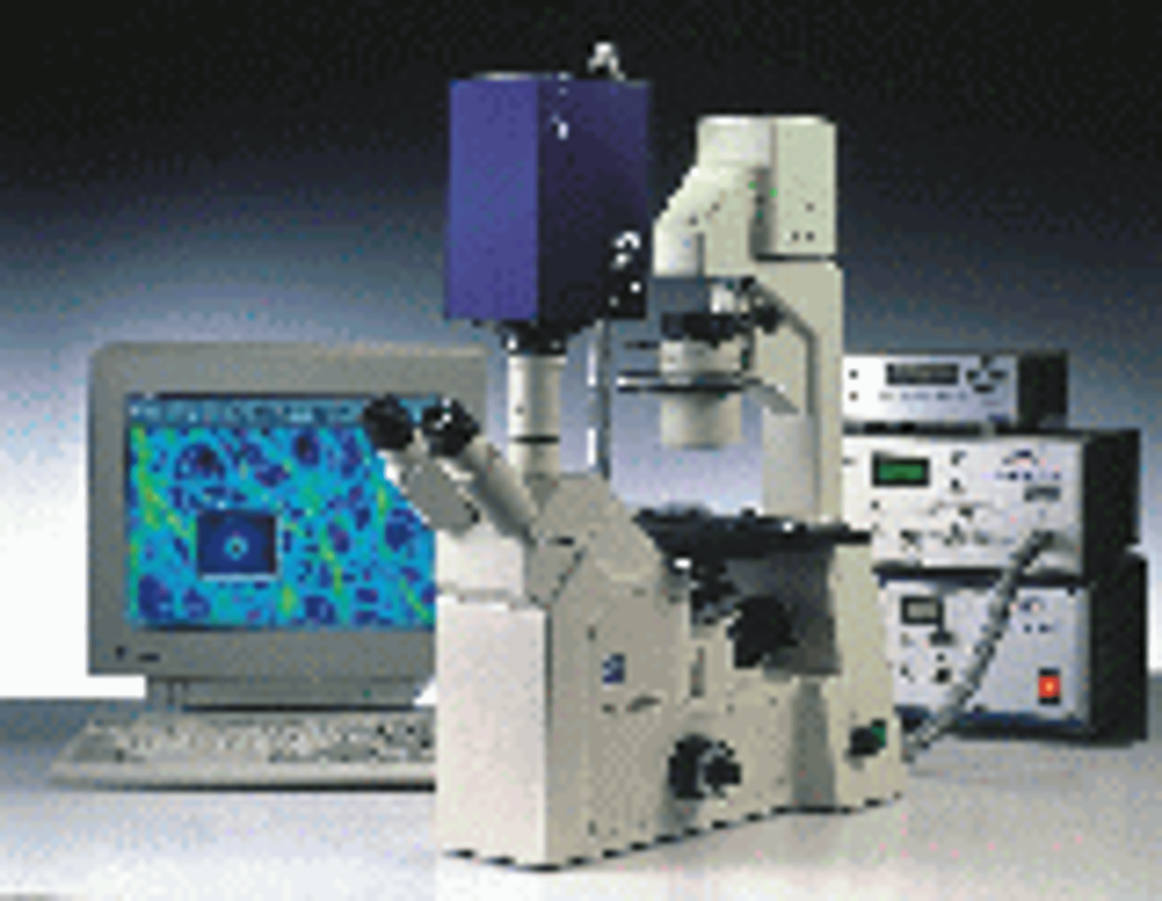
TauScope: Fluorescence Lifetime Imaging Microscopy
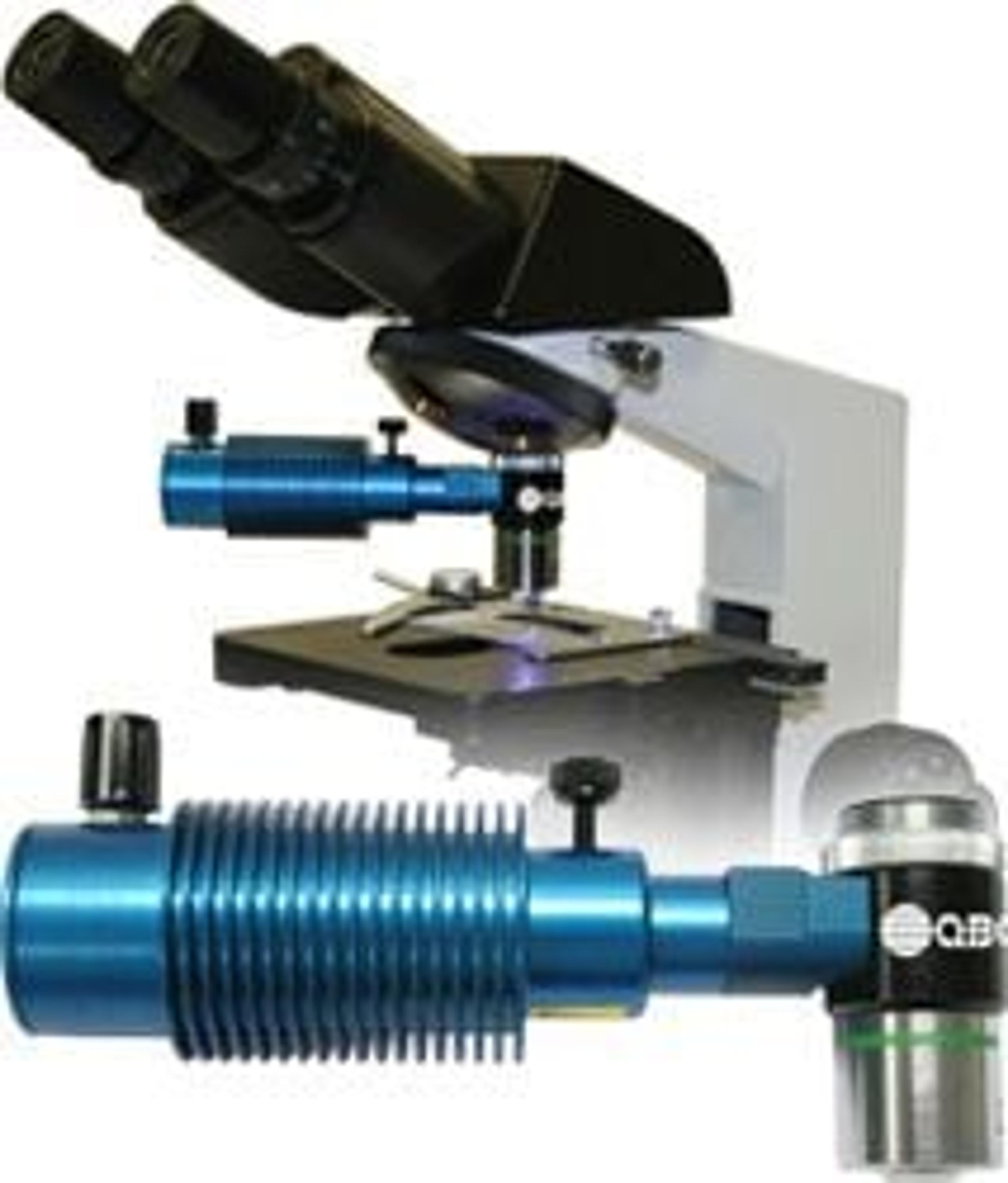
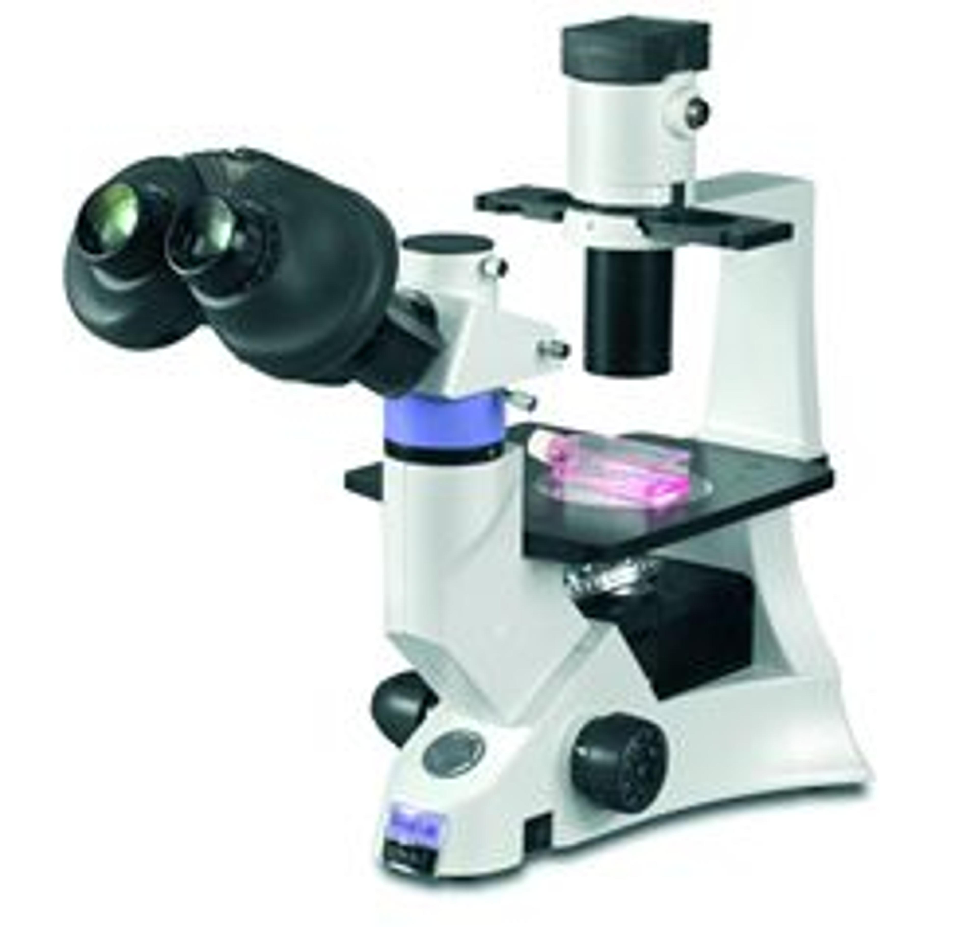
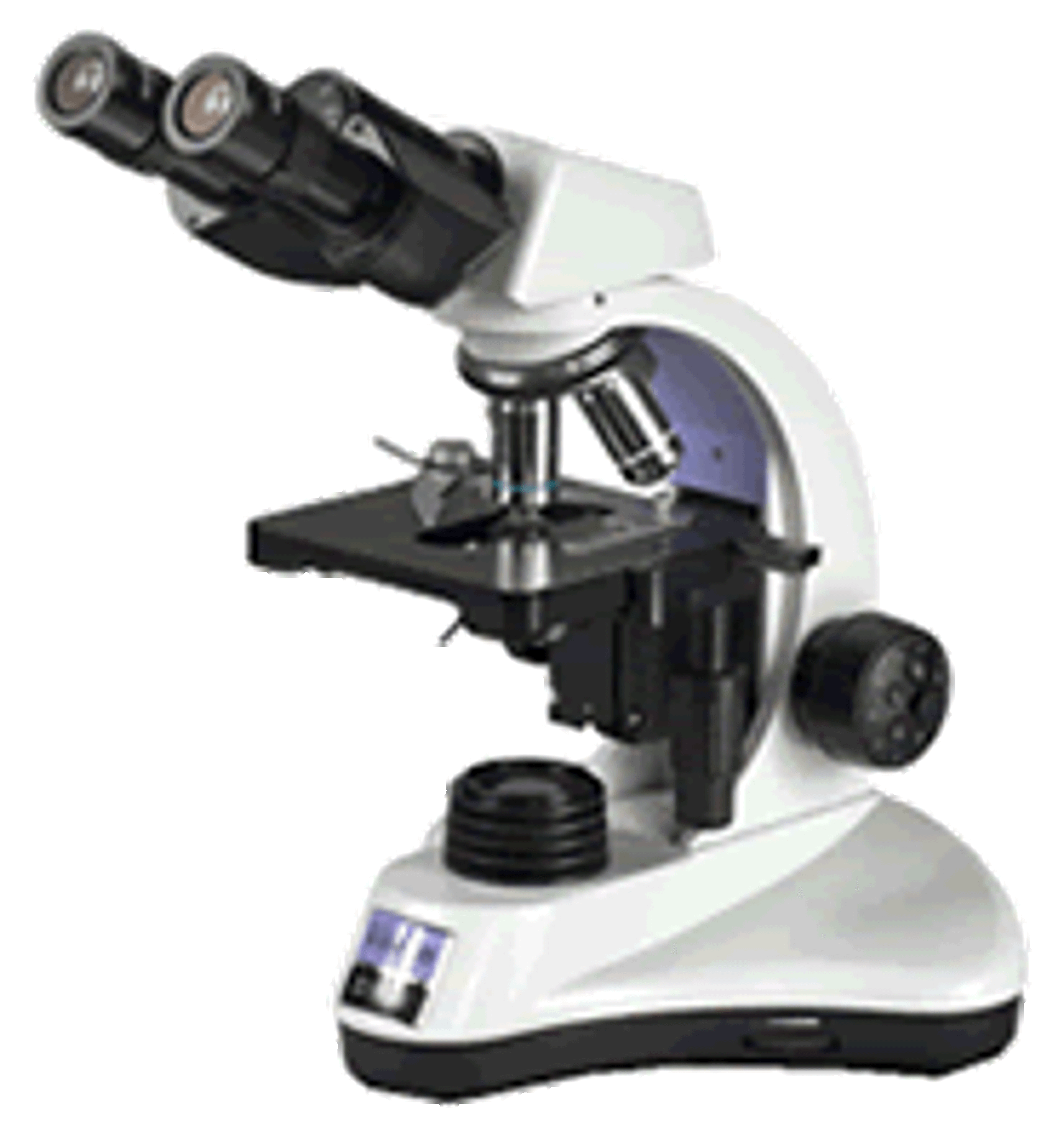
TauTec LLC

TauTec is now offering customized, turn-key time-resolved imaging systems incroporating state-of-the-art intensified, picosecond gated ICCD camera, choice of excitation laser source (mode-locked lasers up to 100 MHz), optical microscope (inverted or upright), imaging spectrograph and the required accessory electronics. The modular system is suitable for various applications such as FLIM (Fluorescence Lifetime Imaging Microscop…

0.0/5.0
|0 Reviews

QBC ParaLens Advance Fluorescent Microscope Attachment

Woodley Equipment Company Ltd

The QBC ParaLens Advance microscope attachment is a unique solution to fluorescence microscopy in that it combines the critical components of an epi-fluorescence microscope, namely the light source and filter set, with a microscope objective in a compact, durable format that can transform virtually any compound light microscope into an epi-fluorescence microscope. Utilising LED technology, the ParaLens light source lamp has a…

0.0/5.0
|0 Reviews


ISOSLIDE® Reticulin Control Slides for the detection of reticular fibers in histology

Merck KGaA, Darmstadt, Germany

To ensure that the test preparations contain the material relevant for diagnostics and the target structures can be visualized by staining with good differentiation, it is recommended to use a control preparation. ISOSLIDE® control slides enable a direct comparison of typical control material with laboratory-internal specimens.Reticular fibers consist of thin bundles of fine fibrils of type III collagen. These collagen fibrils…

0.0/5.0
|0 Reviews


Mantis Eyepieceless Stereo Viewer

Vision Biomed

With in excess of 150,000 installed units worldwide, Mantis has become the low magnification stereo viewer of choice in a vast range of laboratory and industrial applications. All of these applications require bright, well contrasted images that are easy to view, especially for prolonged periods. The patented spatial imaging technology used by Mantis provides true optical 3D images, together with large working distances an…

0.0/5.0
|0 Reviews

DX41 High Magnification Routine Laboratory Microscope

Vision Biomed

The DX41 offers excellent optical performance with a stable compact design and reduced footprint providing ample bench space for accessories. Vision Biomed’s infinity corrected optics provide outstanding contrast and resolution. As a system microscope, Vision Biomed’s DX41 is particularly well suited to clinical and research microscopy applications.  A modular design adds to its versatility with optimised ergonomics making f…

0.0/5.0
|0 Reviews

DX61 Inverted Routine Microscope

Vision Biomed

The DX61 offers life science professionals convenience, performance and flexibility combining infinity corrected optics with innovative mechanical design and is particularly suited to most tissue culture applications. - Outstanding optical performance - Infinity optical system- Ergonomically optimised and compact design- Optional ISIS Expanded-Pupil eyepieces increase eye-reliefVision Biomed have developed the DX61 with the…

0.0/5.0
|0 Reviews

DX21 Routine Microscope

Vision Biomed

The DX21 raises the bar in educational and clinical examination. The infinity optical system provides clear sharp images and Vision Biomed ’s ergonomic design ensures comfortable viewing over extended periods of use.- Superb ergonomics for optimum operator comfort- Outstanding optical performance - Modular design allows for easy addition of accessories - Compact stable design The DX21 is a robust, simple to use, purpose built…

0.0/5.0
|0 Reviews

Expanded Pupil Technology

Vision Biomed

Isis expanded pupil is newly patented multi-lenticular technology to a new device designed to enhance the ergonomics of all conventional binocular microscopes. Vision’s Isis accessory fits directly into the microscope’s eyepiece tubes to deliver Expanded Pupil technology, offering an exit ray bundle twelve times larger than a traditional eyepiece.Expanded Pupil technology allows a much greater freedom of operator head movement…

0.0/5.0
|0 Reviews

Morphology Explorer V2 Image Analysis BioApplication

Morphology Explorer V2 is a powerful general-purpose image analysis application for measuring size, shape, and distribution of cells, colonies, and subcellular components.  It performs sophisticated calculations in a turn-key, automated manner allowing higher throughput of complex cell-based assays.  This application has been biologically validated, enhancing confidence in your image-based data.    Available for the Cellomics…

0.0/5.0
|0 Reviews

Cell Health Profiling V2 Image Analysis BioApplication for Apoptosis and Cytotoxicity

Cell Health Profiling V2 is a flexible image analysis application employing advanced cell-level logic to detect and classify cell health-related events.  It analyzes patterns of cellular fluorescent labeling that can be used to classify cells as undergoing apoptotic, necrotic, or other cell death mechanisms.  This application has been biologically validated, enhancing confidence in your image-based data.  Available for the Cel…

0.0/5.0
|0 Reviews

Cell Cycle V2 Image Analysis BioApplication

The Cell Cycle V2 BioApplication is a sophisticated image analysis algorithm that performs automated analysis of the cell cycle phase of individual and populations of cells.  It reports cell cycle phase as well as the state of up to 3 target proteins for every cell analyzed, and correlates the target data with DNA content.  This application has been biologically validated, enhancing confidence in your image-based data.  Availa…

0.0/5.0
|0 Reviews

Cytotoxicity V2 Image Analysis BioApplication

The Cytotoxicity V2 BioApplication is an image analysis application that simultaneously measures four markers of cytotoxicity (nuclear fragmentation/condensation, permeability, lysosomal mass-pH, and cell count) and reports individual and composite indicies of cytotoxicity.  The V2 version has been enhanced with object segmentation to allow clumped cells to be efficiently analyzed.  Available for the Cellomics ArrayScan VTI, A…

0.0/5.0
|0 Reviews

Extended Neurite Outgrowth V2 Image Analysis BioApplication for Neurobiology

Extended Neurite Outgrowth V2 is an advanced image analysis algorithm that can quantitate neurite outgrowth with features such as cell body area and branch point quantitation, cell subpopulation analysis, Ability to analyze neuron subsets expressing up to two distinct neurotransmitters.  It reports all features at the cell, well, and subpoulation level, and is biologically validated to enhance confidence in your image-based da…

0.0/5.0
|0 Reviews

Compartmental Analysis V2 Image Analysis BioApplication

Compartmental Analysis V2 is a powerful general image analysis tool for quantitating intracellular intensity changes in specific sub-cellular regions across multiple fluorescent channels.  It performs spot analysis and reports intensity ratios and differences among sub-cellular regions and/or different fluorophores.  This application has been biologically validated, enhancing confidence in your image-based data.  Available for…

0.0/5.0
|0 Reviews

Cell Motility V2 Image Analysis BioApplication for Cancer Research

The Cell Motility V2 BioApplication is a advanced image analysis algorithm for measuring the overall motility of cells.  It quantitates total distance traveled by each cell exposed to growth factor or compound for a quantiative assessment of cell motility.  The V2 version includes advanced analytical capabilities such as population analysis and object segmentation.  This application has been biologically validated, enhancing c…

0.0/5.0
|0 Reviews

Spot Detector V2 Image Analysis BioApplication

Spot Detector V2 performs measurements of number, size, and other features of spots in cells.  It is an easy-to-use and essential component of any image analysis software suite.  This tool is excellent for measuring receptor internalization and trafficking, cell proliferation and counting, Analyzing populations. This application has been biologically validated, enhancing confidence in your image-based data.  Available for the…

0.0/5.0
|0 Reviews

Neurite Outgrowth V2 Image Analysis BioApplication for Neurobiology

The Neurite Outgrowth V2 BioApplication is a streamlined image analysis algorithm for morphological analysis of neurite outgrowth. Utilizes fluorescent images of neurons and neurites stained for tubulin to quantitate the number and length of neurites in a population of cells.  This application has been biologically validated, enhancing confidence in your image-based data.  Available for the Cellomics ArrayScan VTI, ArrayScan x…

0.0/5.0
|0 Reviews

Cyto-Cell Membrane Translocation V2 Image Analysis BioApplication

Cyto-Cell Membrane Translocation V2 is a multi-channel image analysis algorithm for analysis of targets that translocate from cytoplasm to cell membrane.  It utilizes colocalization of fluorescent markers to report whether the target is co-localized to the membrane, and reports cells' behavior relative to a control population.   This application has been biologically validated, enhancing confidence in your image-based data.  A…

0.0/5.0
|0 Reviews

GPCR Signaling V2 Image Analysis BioApplication

GPCR Signaling V2 is a high-content image analysis application for quantitation of the activation of G-protein coupled receptors.  Classifies cells into different phases based on pattern of receptor complex distribution and reports details of and percentage of cells with Type A and Type B recycling activity.  This application has been biologically validated, enhancing confidence in your image-based data.  Available for the Cel…

0.0/5.0
|0 Reviews