Invitrogen™ Annexin V Alexa Fluor™ 488 Ready Flow Conjugate
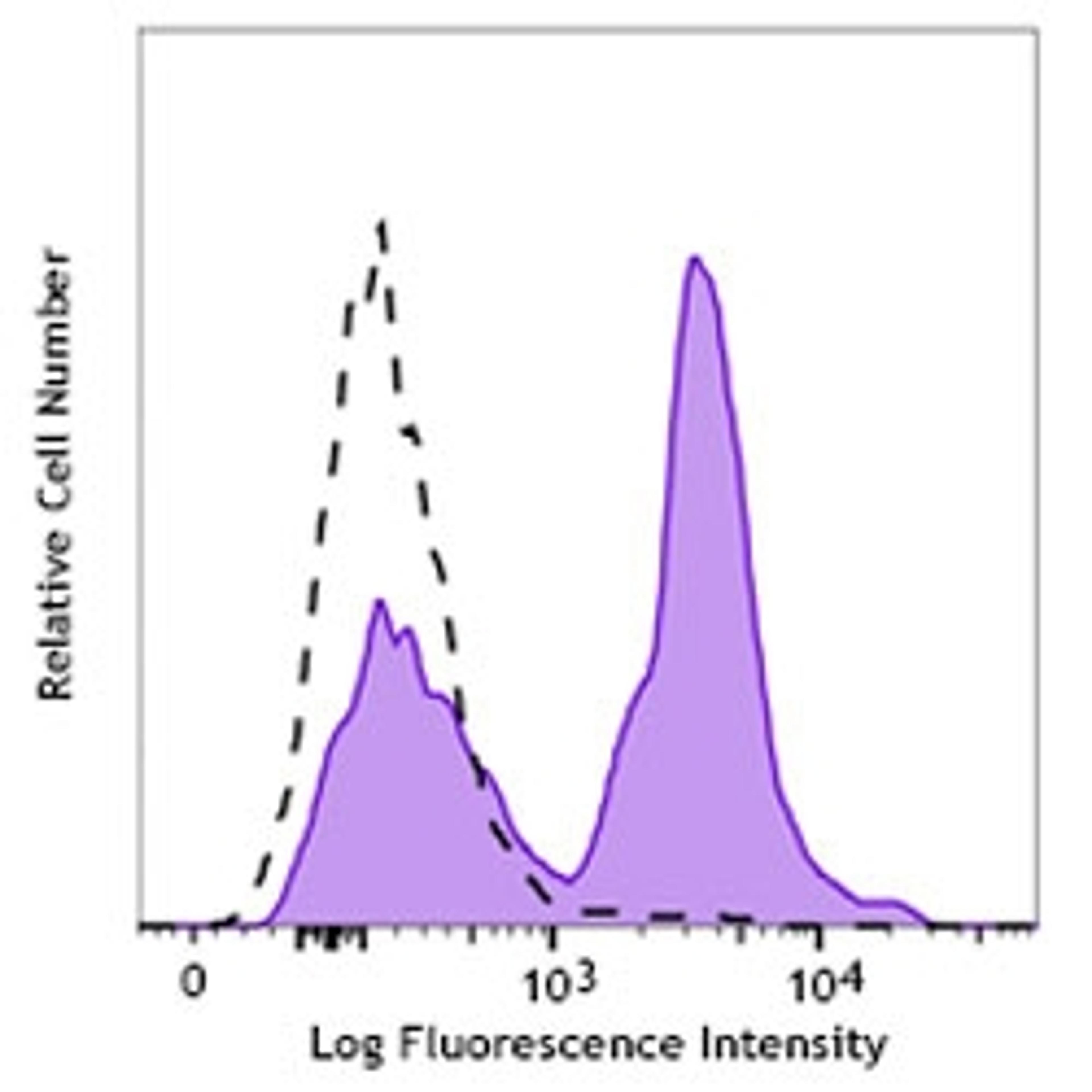
Thermo Fisher ScientificAnnexin V Alexa Fluor 488 Ready Flow Conjugate is a bright, easy-to-use stain for apoptosis with high affinity for phosphatidylserine (PS), which becomes exposed on the outer leaflet of cells undergoing apoptosis. The Annexin V Alex Fluor 488 conjugate is excitable at 495 nm with an emission peak at 519 nm.FeaturesStable at room temperatureConvenient, ready-to-use format—no need to dilute, weigh, or pipetteCan be added directl…