HALO AI
Neural network driven tissue and cell classification for research & routine pathology
Currently the best image viewer and analysis software on the market that I've used.
Image analysis
Currently the best image viewer and analysis software on the market that I've used.
Review Date: 8 Jun 2022
wonderful products, it is far beyond our expectations.
analyze tumor sections
Our lab both have the Halo Image Analysis System and the Halo AI, honestly speaking, the Halo system is beyond our expectations, especially the analysis process, annotation tools, the adjustable parameters, the texture and morphological recognition of nuclear and specific structure, and the after-sale care I strongly recommend you guys to choose it. it really worth the money.
Review Date: 7 Jun 2022
Great results, can't live without this instrument!
Analysis in tissue figure
Scientists can achieve lots of high-quality analysis data about figures by HALO software. Many functions of HALO are very helpful to study images, such as TMA, spatial analysis, high-Plex FL, and especially HALO AI. It is easy to operate. Its interface is simple, friendly, and convenient. Once we have problems, a Halo technician is able to help us to solve them in time.
Review Date: 1 Jun 2022
Great result. Nice tool for translational pathology research.
RNAscope and IHC analysis
We are interested in applying HALO AI in RNAscope and IHC analysis. Thanks to Yongtian ZHAO to give us wonderful training and support. We are improving and confident to apply it in the RNAscope assay development and scoring.
Review Date: 1 Jun 2022
Great
Halo for image research
Quick to use
Review Date: 1 Jun 2022
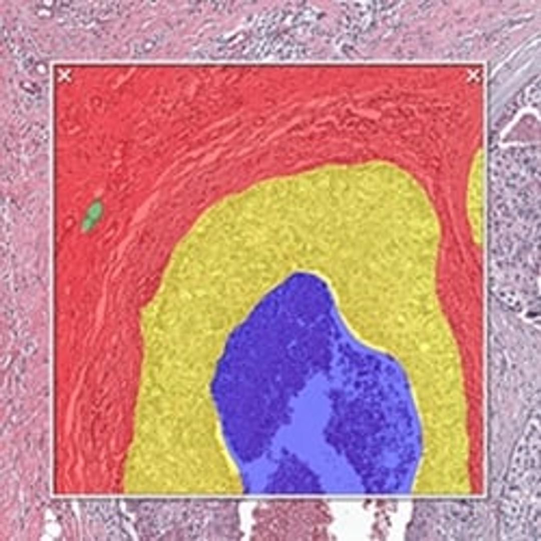
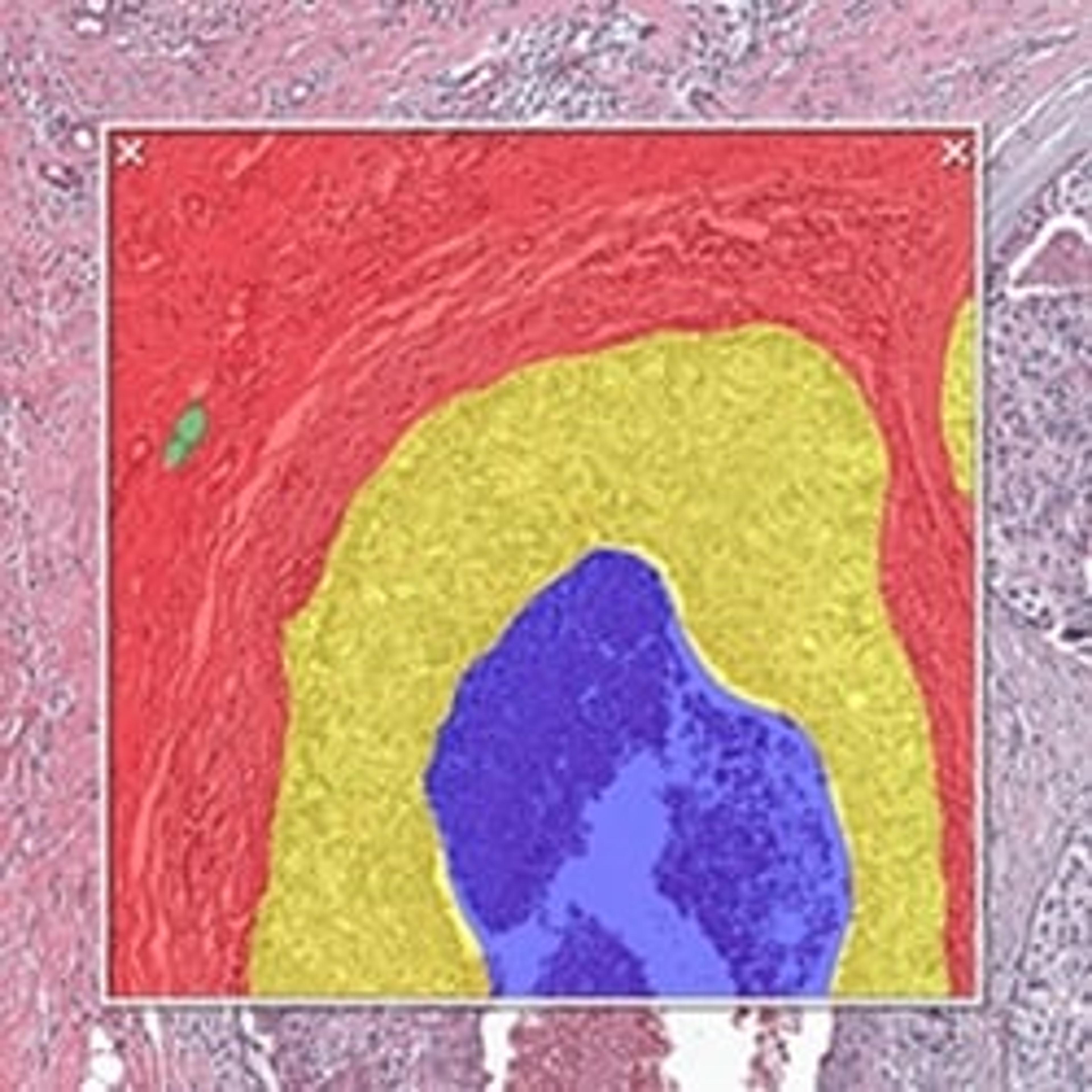
HALO AI is a collection of train-by-example classification and segmentation tools underpinned by advanced deep learning neural network algorithms. HALO AI classifiers can be trained to quantify tissue classes, to segment tissue classes for analysis with other HALO image analysis modules, to find rare events or cells in tissues, and to categorize cell populations into specific phenotypes.
SIMPLE & INTUITIVE WORKFLOW
HALO AI is fully integrated with the intuitive, easy-to-use HALO and HALO Link viewers and employs a simple three-step workflow. After defining what tissue classes or cell phenotypes you would like to segment, you train the neural network by drawing annotations – no computer programming or AI knowledge required. Trained classifiers can be applied to segment tissue and cells on any whole slide image or region of interest.
POWERFUL TISSUE SEGMENTATION
HALO AI now includes the option of three powerful neural networks – VGG, DenseNet and MiniNet. VGG, a well-known and more traditional network, was used to build the Indica Labs submission in the CAMELYON17 challenge and was the first neural network integrated with HALO AI. DenseNet is a more modern network capable of creating more robust classifiers at higher resolution compared to VGG. MiniNet, a custom network developed at Indica Labs, is more shallow than VGG or DenseNet, but can produce a solution quickly with limited training data and is therefore useful for testing new AI applications.
EXCEPTIONAL CELL CLASSIFICATION
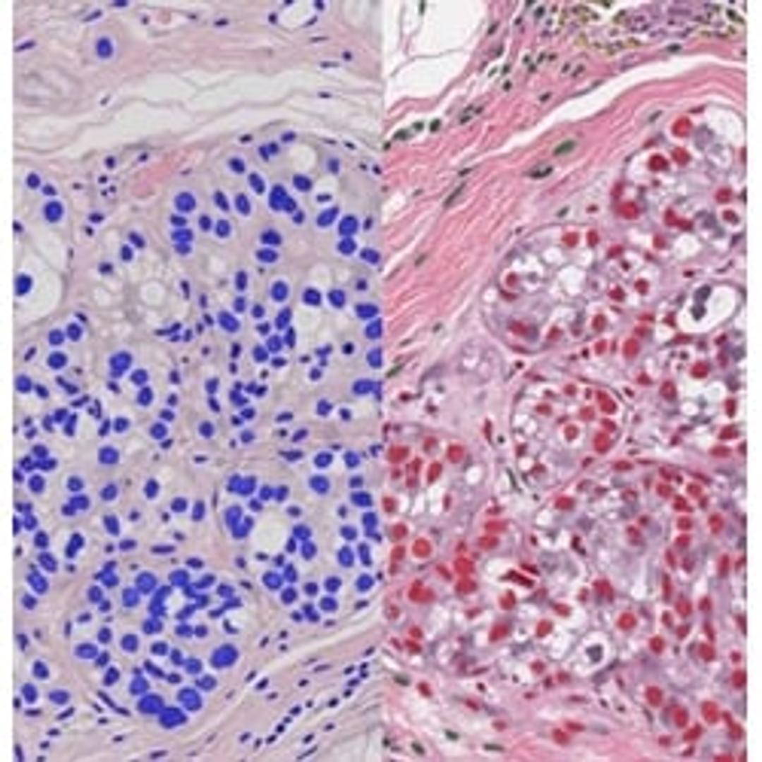
Segment nuclei with the new Segmentation classifier. Utilize HALO AI’s pretrained networks for H&E, single IHC, or DAPI stained images for an out of the box solution. Or train your own nuclei segmentation network for a specific application (unique tissue or advanced staining protocols). Once nuclei are segmented, take it a step further using the Nuclei Phenotyper classifier to automatically assign cells into user defined phenotypes with a few quick training examples.

