Light Microscopy Products & Reviews
25
Light microscopes or optical microscopes are used to visualize microscale objects under magnification, including cells, clinical specimens and materials. Lab equipment for light microscopy includes confocal microscopes, fluorescence microscopes, zoom and stereo microscopes. Microscope slides and imaging reagents are available for visualizing samples, as well as various microscope stages and incubators for large or temperature-sensitive samples. Find the best light microscopes in our peer-reviewed product directory: compare products, check customer reviews and receive pricing direct from manufacturers.
Selected Filters:
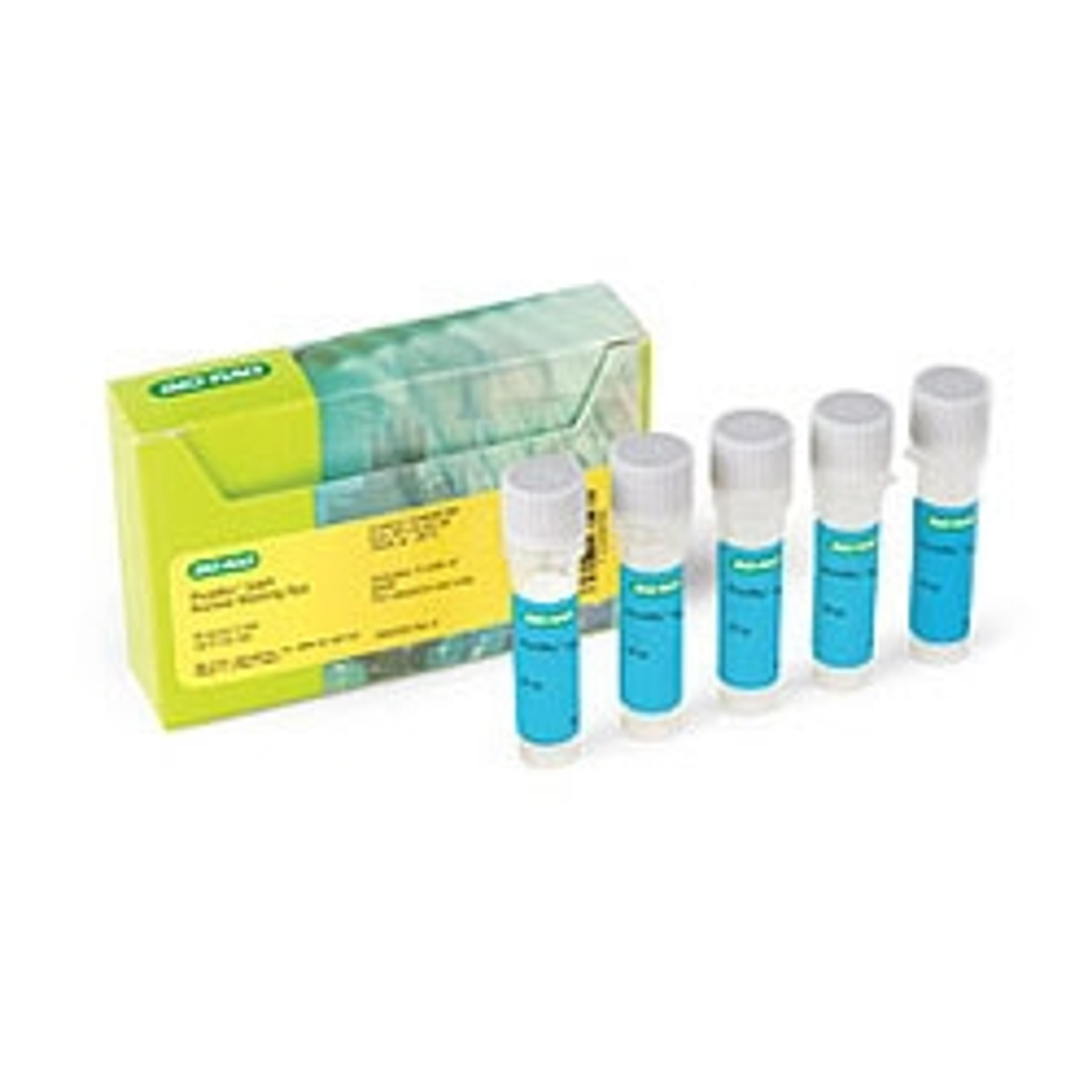
PureBlu™ DAPI Nuclear Staining Dye
Bio-RadPureBlu™ Nuclear Staining Dyes are designed to specifically stain the nuclei of cells in fixed and unfixed samples for fluorescence microscopy and cell imaging applications.
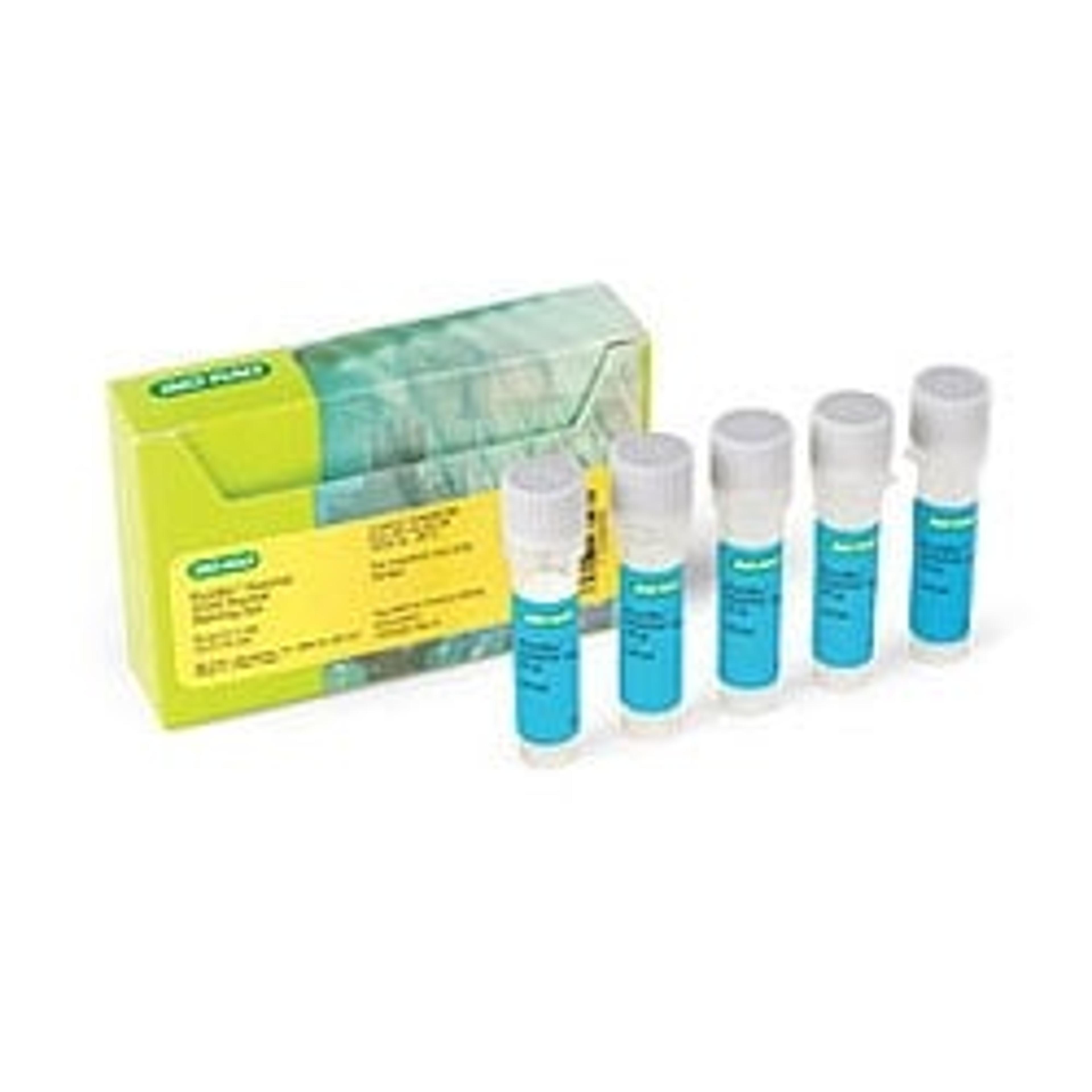
PureBlu™ Hoechst 33342 Nuclear Staining Dye
Bio-RadPureBlu™ Nuclear Staining Dyes are designed to specifically stain the nuclei of cells in fixed and unfixed samples for fluorescence microscopy and cell imaging applications.
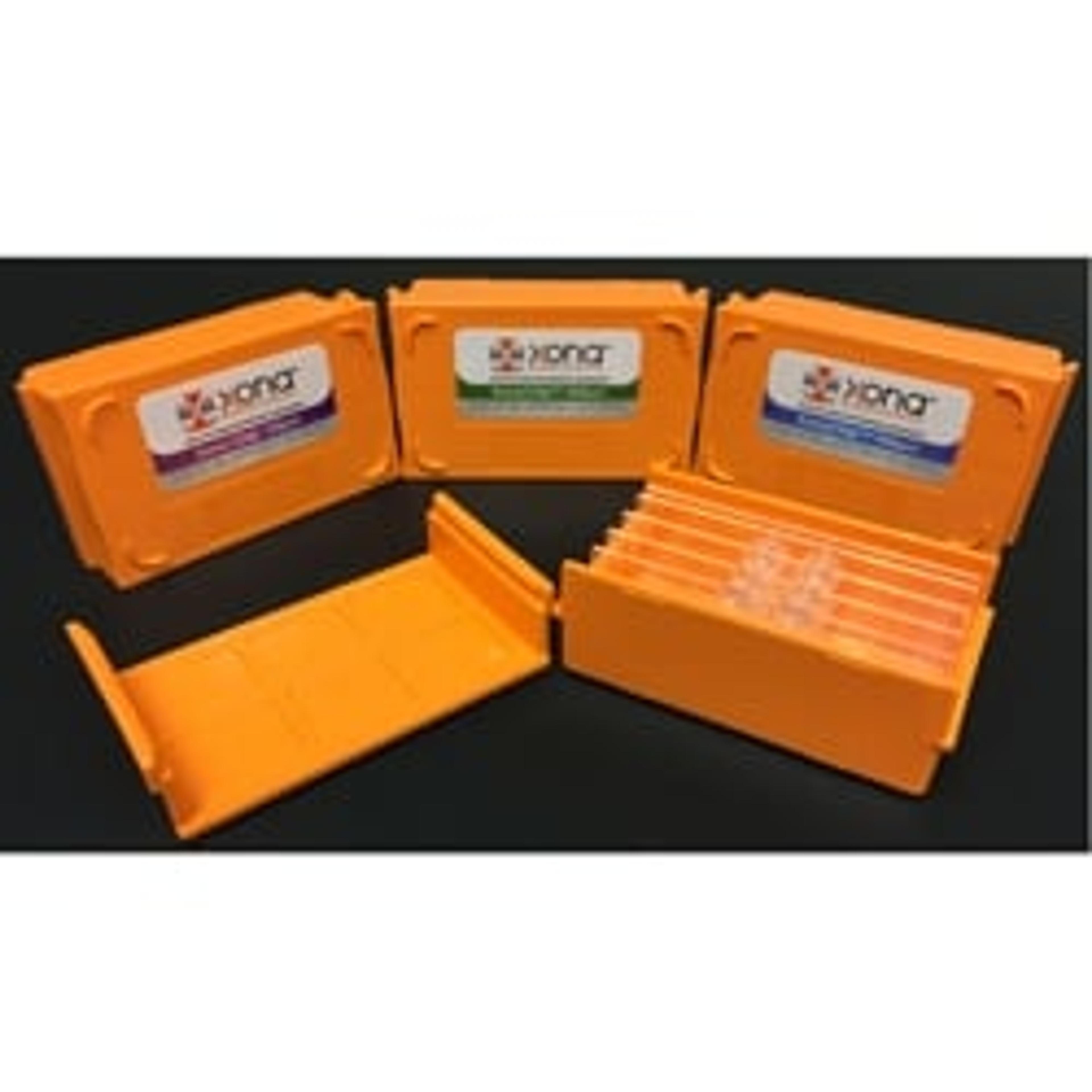
XonaChips™
Xona MicrofluidicsXonaChips™ are the latest innovation in neuron culturing! XonaChips™ are pre-assembled and pre-bonded, making them easier-to-use than our original silicone devices. These plastic chips improve long-term culture of animal model neurons. They are especially useful for culturing human stem cell-derived neurons, providing better cell attachment and long-term growth. XonaChips™ are optically transparent and ideal for fluorescence…
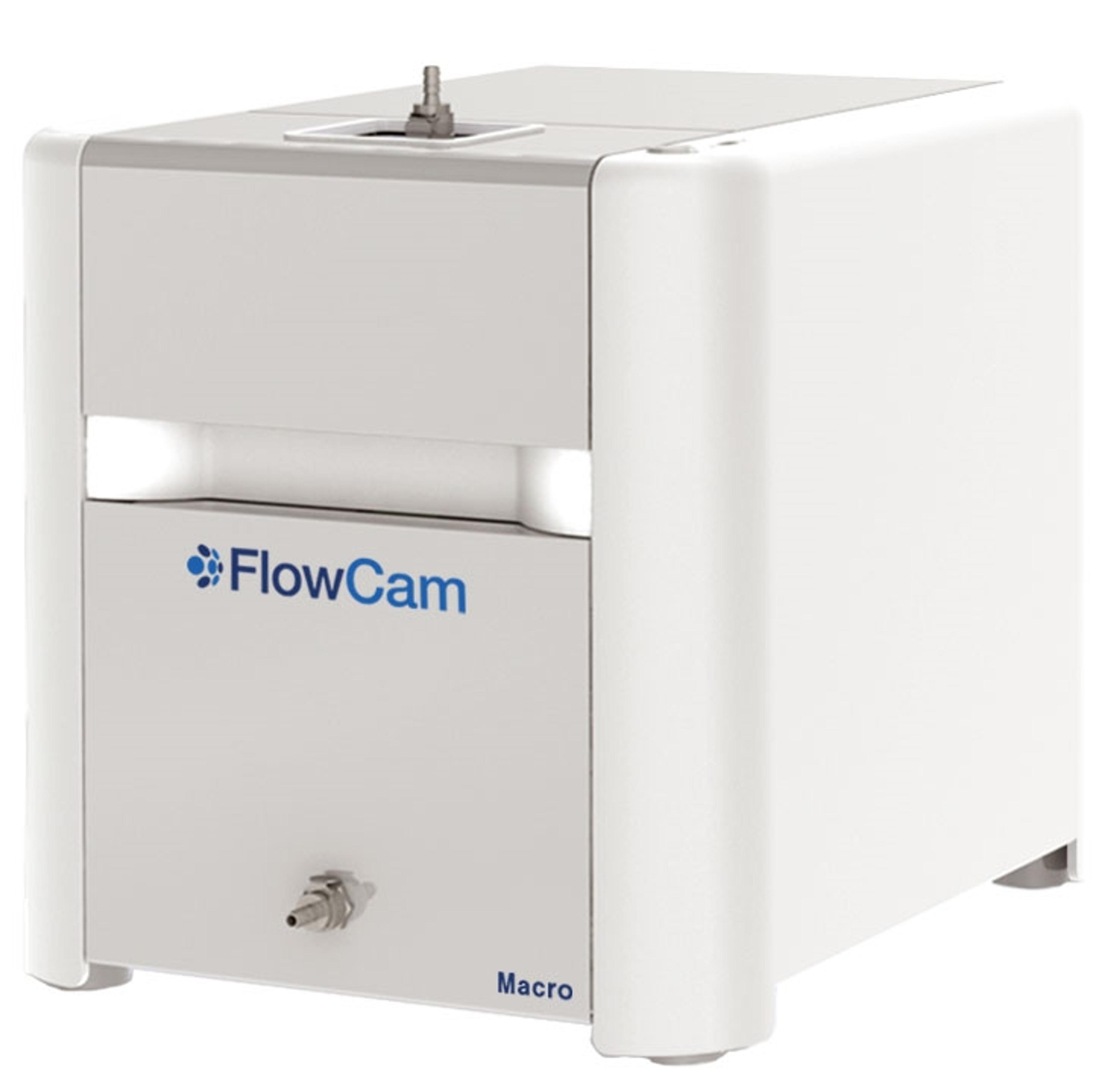
FlowCam Macro
Yokogawa Fluid Imaging Technologies, IncExtend your particle imaging capabilities from 300 μm to 5 mm with FlowCam® Macro for aquatic and environmental research and materials characterization. Obtain detailed morphological data along with accurate counting and sizing measurements to enable differentiation of diverse zooplankton species and particle types.
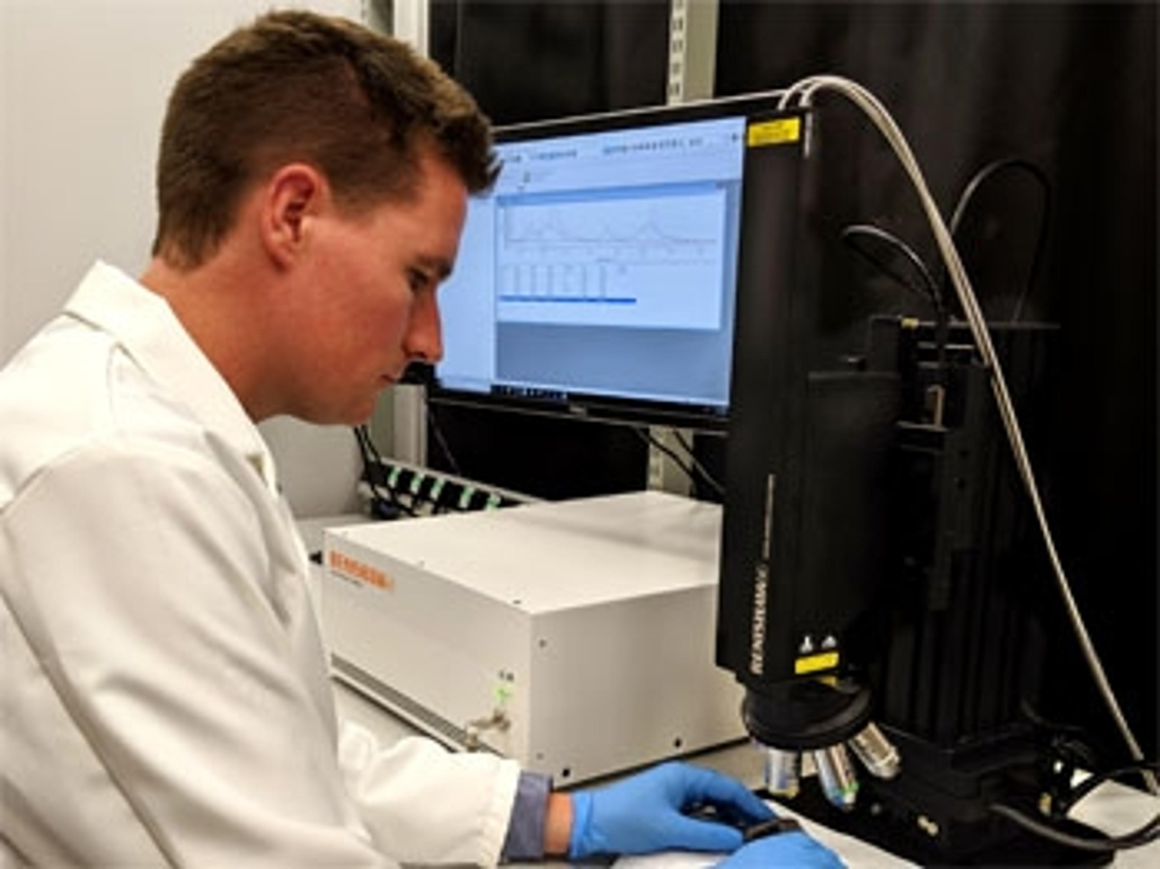
Virsa™ Raman Analyser
Renishaw plc.Use the Virsa Raman Analyser, Renishaw's latest high-performance Raman spectroscopy system, to take your spectroscopic analysis away from the confines of the laboratory microscope to new samples and environments.
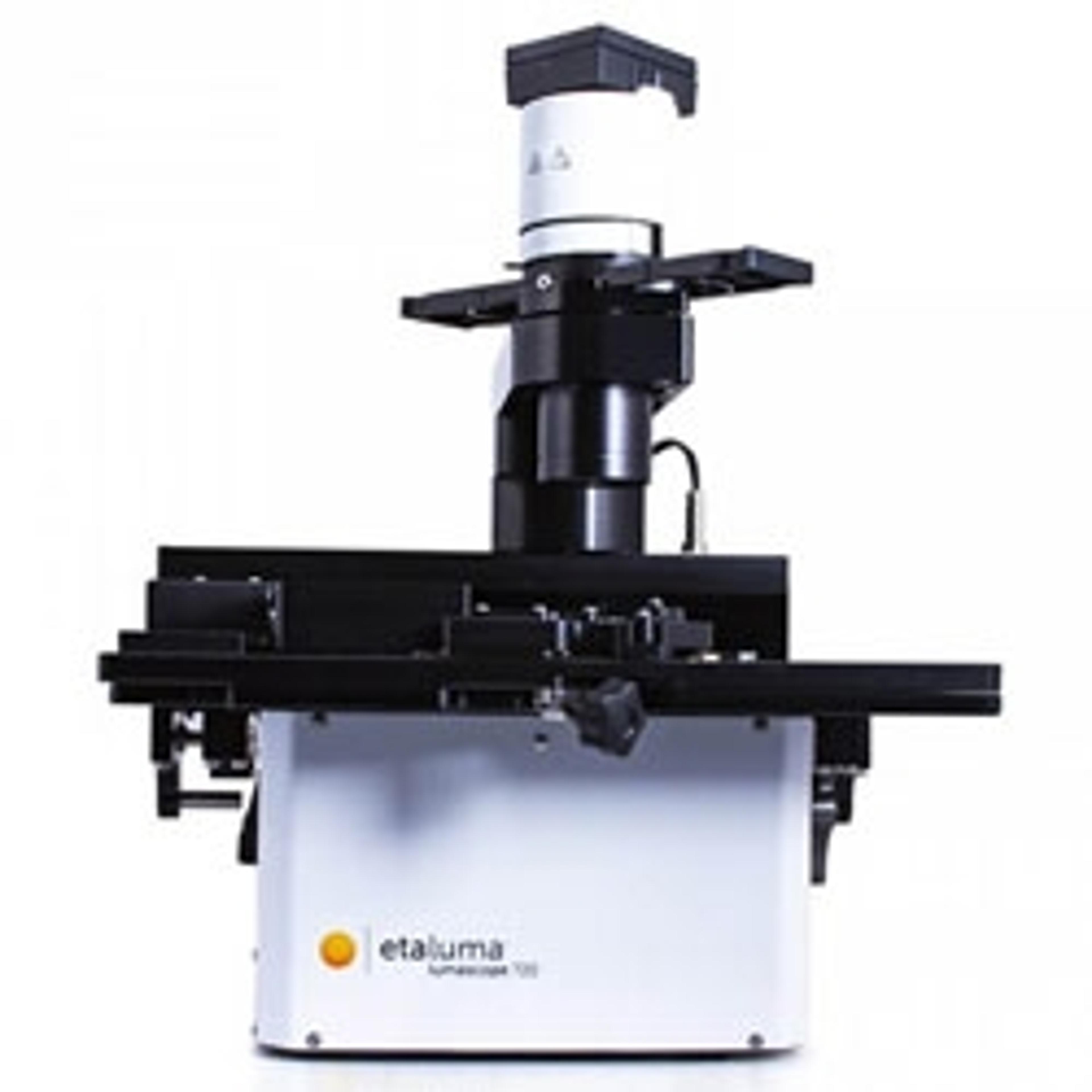
Etaluma Lumascope Range
Labtech InternationalThe Etaluma Lumascope range provides a unique approach to live cell imaging. These compact and durable systems offer high quality brightfield, phase-contrast and fluorescence imaging. Lumascopes are engineered to withstand the most demanding environments including incubators, hoods and workstations.
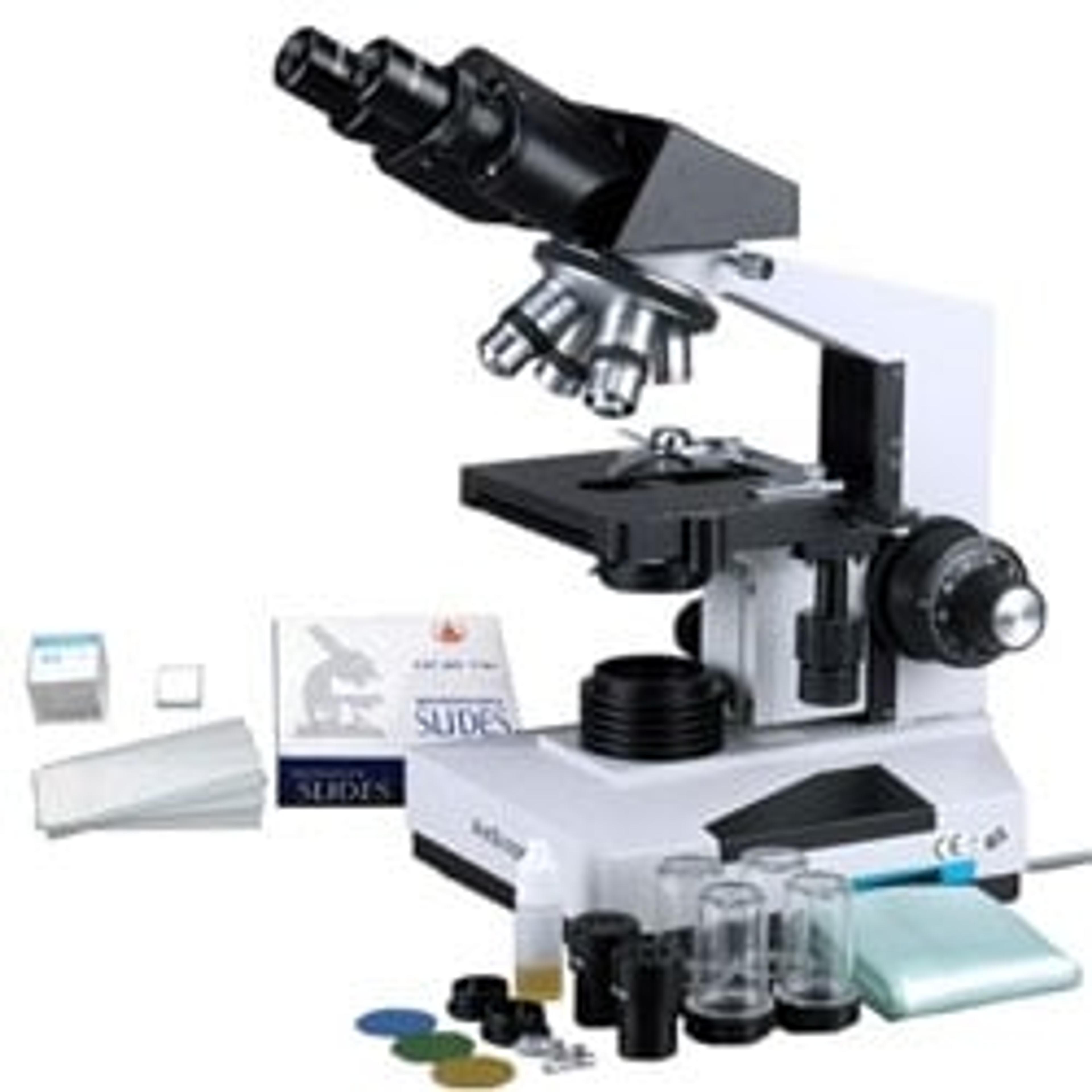
1600x Professional Biological Microscope + 50 Slides + 100 Coverslips
AmscopeThis is a professional binocular microscope with 50pc slides and 100pc coverslips. It is designed for teaching demonstration, clinical examination and research purpose. It comes with a 30 degree inclined 360 degree swiveling compensation free binocular head, 3D mechanical stage and an intensity-variable halogen illumination system. This microscope offers eight levels of magnification, 40X, 64X, 100X, 160X, 400X, 640X, 1000…
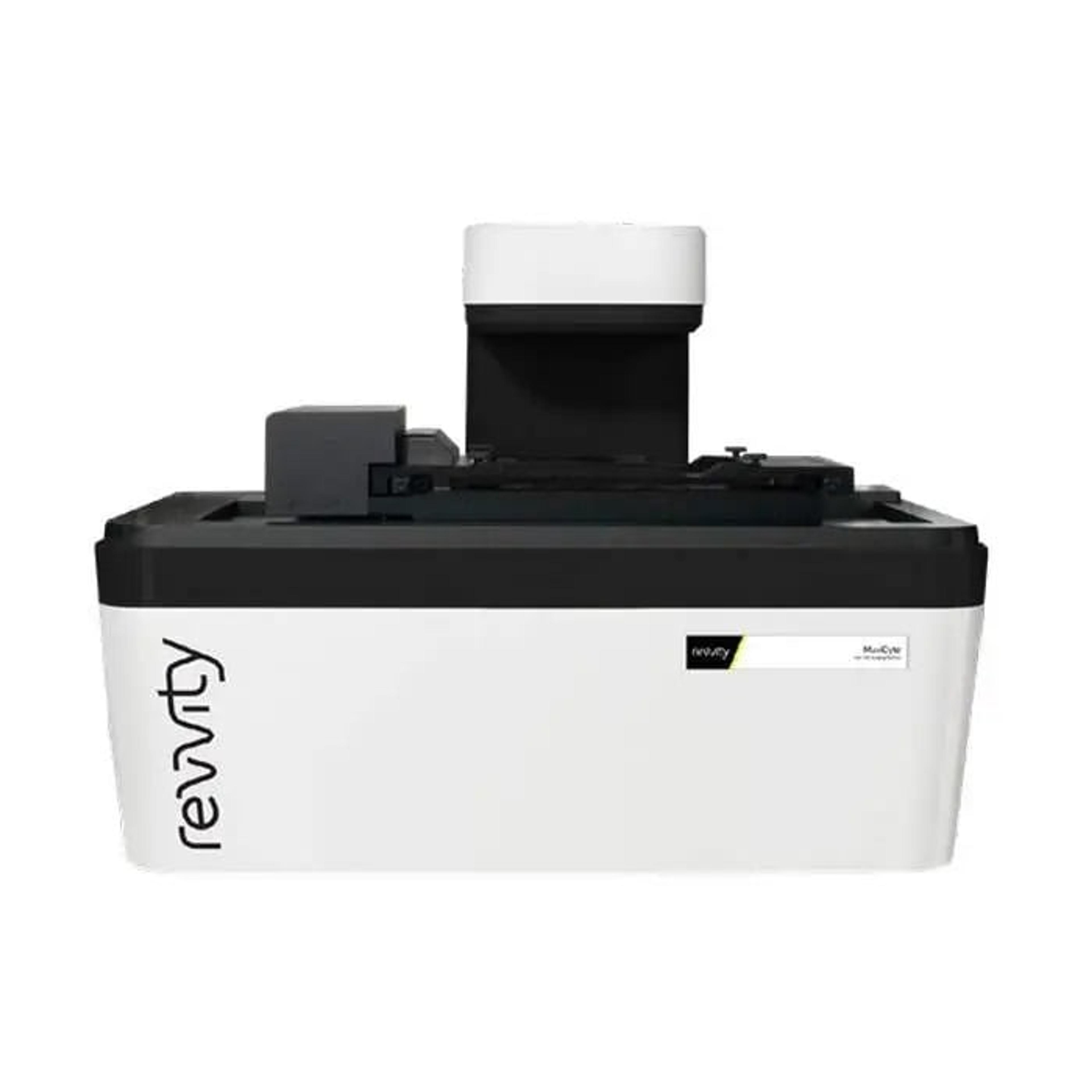
MuviCyte Live-Cell Imaging Kit
RevvityThe MuviCyte™ live-cell imaging system is designed to operate inside your cell-culture incubator, enabling you to maintain your cells under optimal conditions and perform a wide range of assays in a variety of culture vessels.

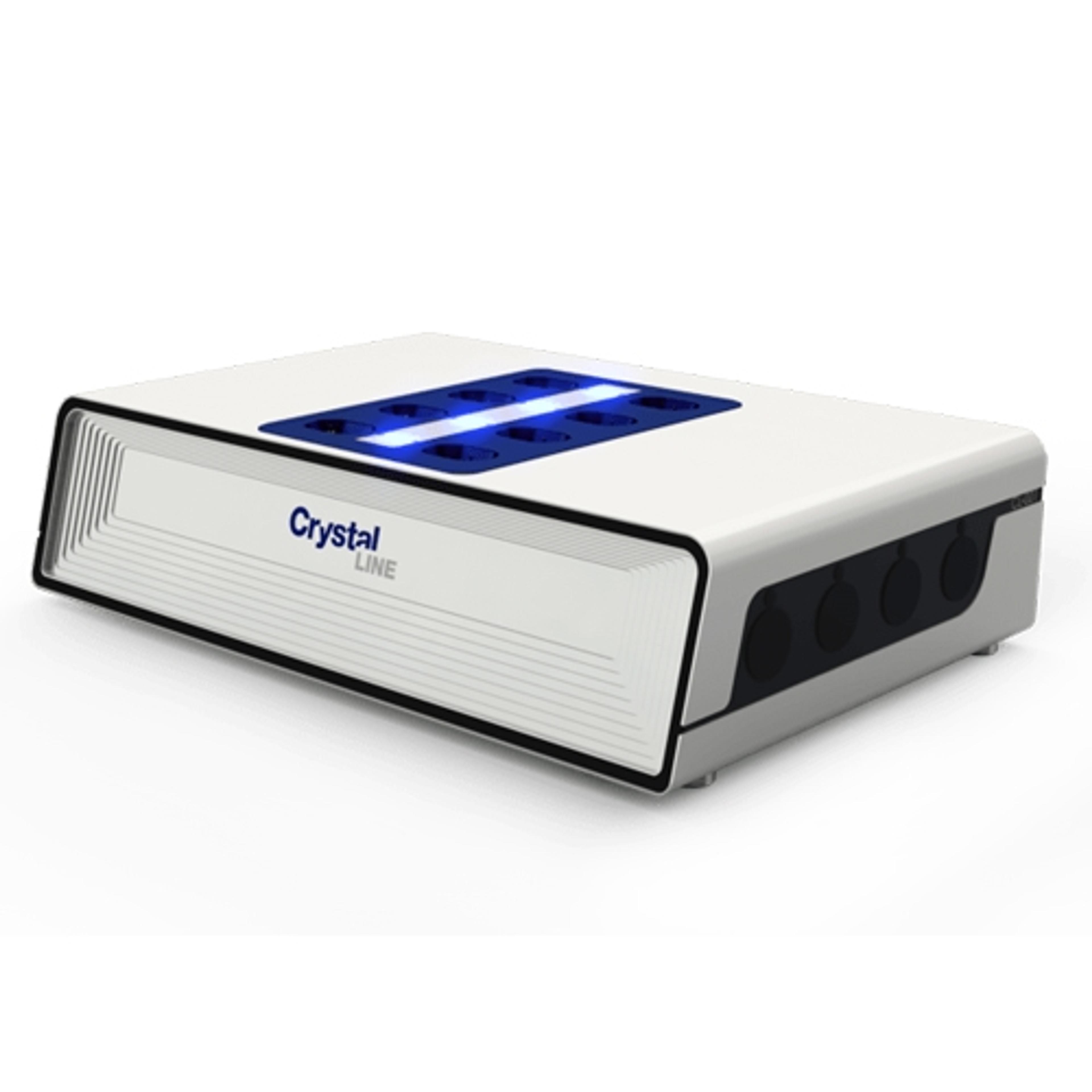
Crystalline PV/RR
Technobis Crystallization SystemsAccess crystallization and formulation information at mL scale with the new Crystalline PV/RR
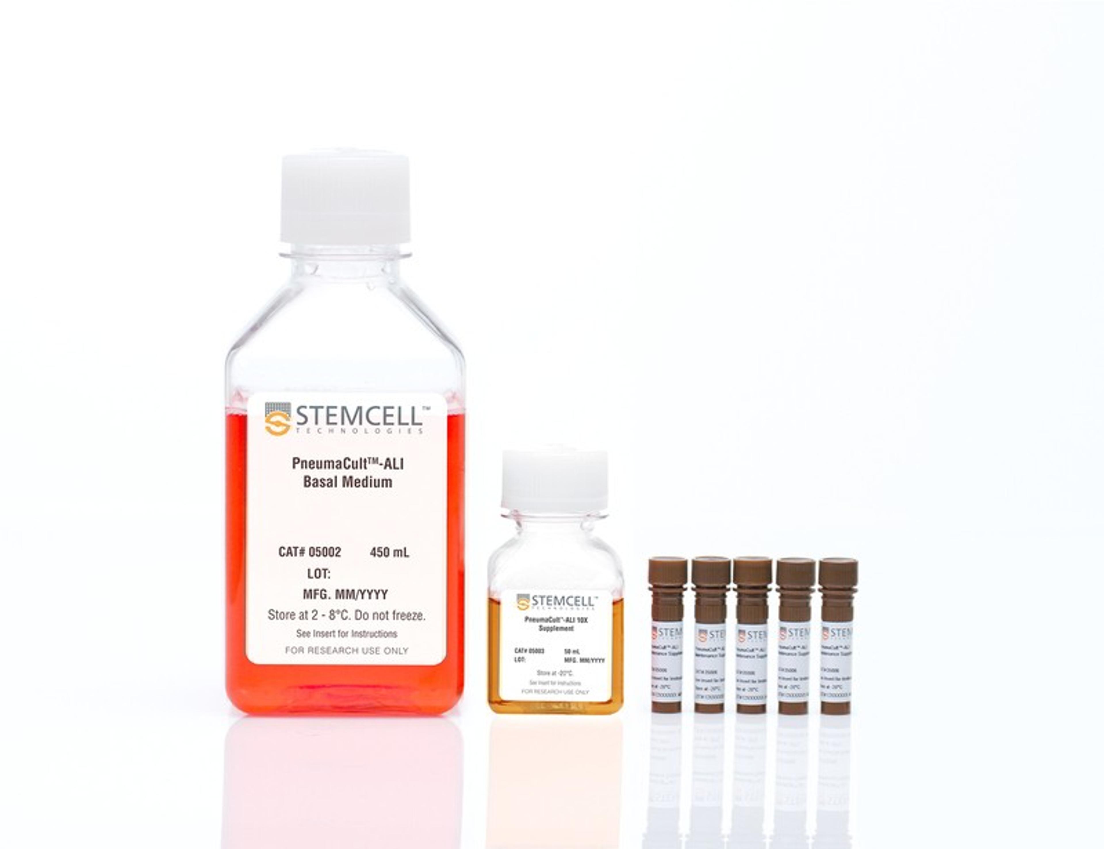
PneumaCult™-ALI Medium
STEMCELL Technologies Inc.Serum- and BPE-free medium for human airway epithelial cells cultured at the air-liquid interface or as airway organoids
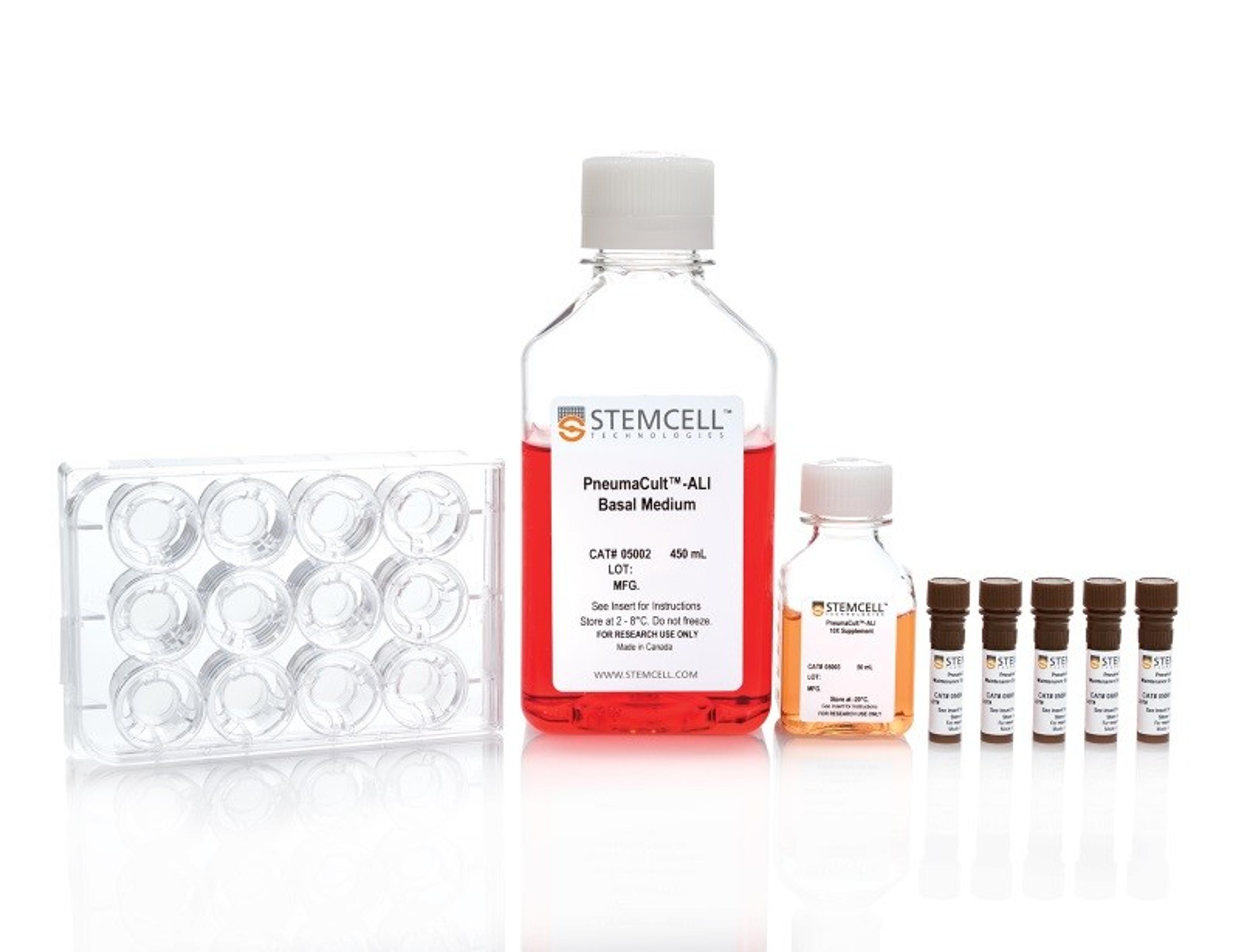
PneumaCult™-ALI Medium with 12 mm Transwell® Inserts
STEMCELL Technologies Inc.Serum- and BPE-free medium for human airway epithelial cells cultured at the air-liquid interface or as airway organoids

PneumaCult™-ALI Medium with 6.5 mm Transwell® Inserts
STEMCELL Technologies Inc.Serum- and BPE-free medium for human airway epithelial cells cultured at the air-liquid interface or as airway organoids

PneumaCult™-Ex Plus Medium
STEMCELL Technologies Inc.Serum- and BPE-free medium for expansion of primary human airway epithelial cells
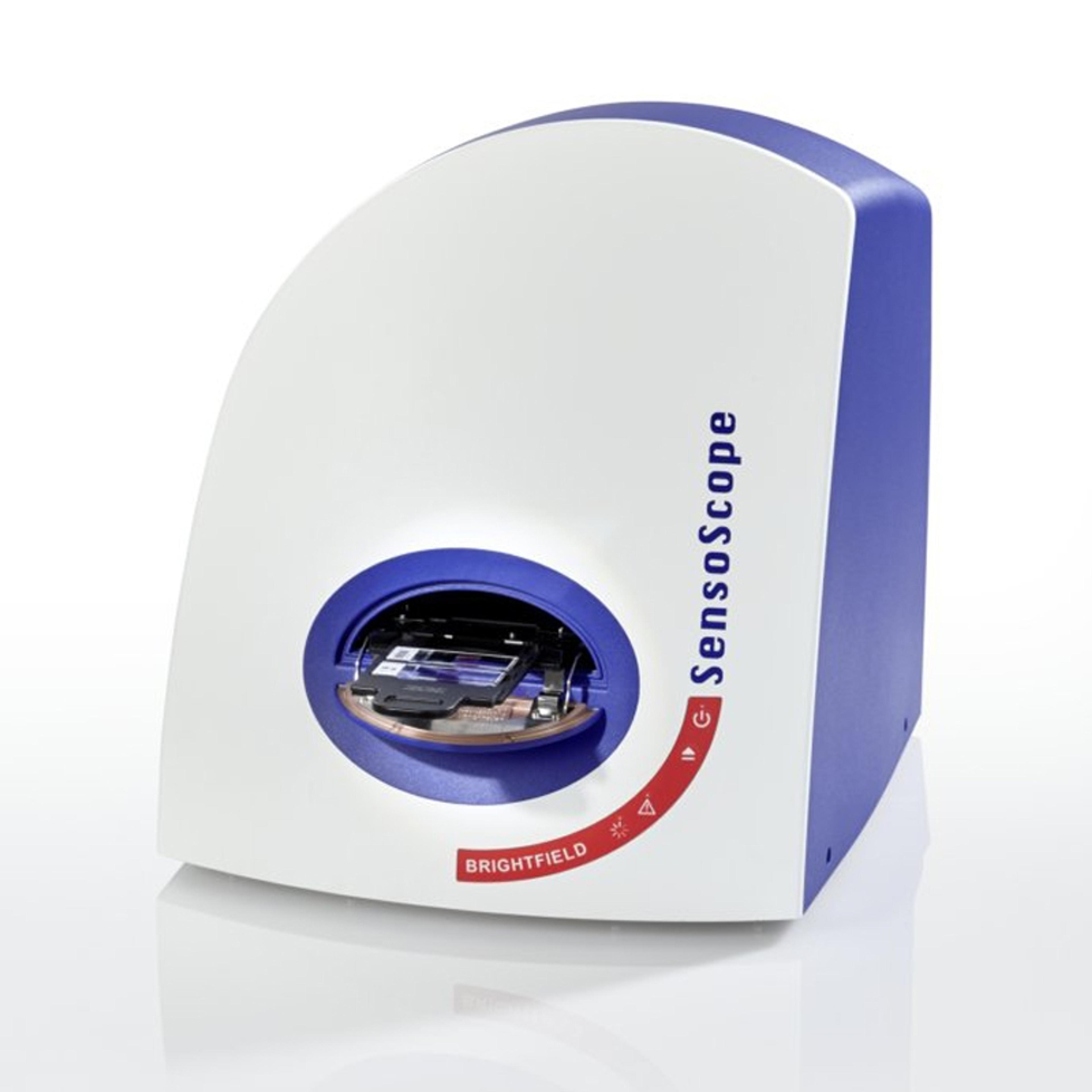
SensoScope® Brightfield
Miltenyi Imaging GmbHPremium digital microscope for live imaging, scanning and real-time diagnostics of multiple slides
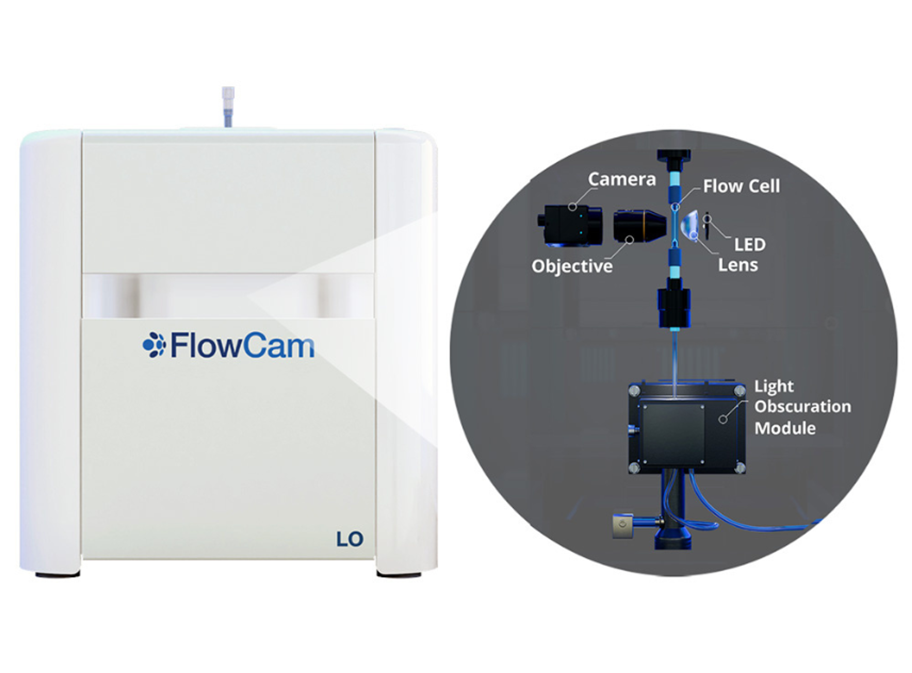
FlowCam LO
Yokogawa Fluid Imaging Technologies, IncInnovative particle characterization with FlowCam® LO combines flow imaging microscopy (FIM) and light obscuration (LO) into a single analytical solution. Beyond the compendial light obscuration method to fulfill USP <787> and <788> requirements, flow imaging microscopy provides an orthogonal method for quality control of subvisible particulate matter.
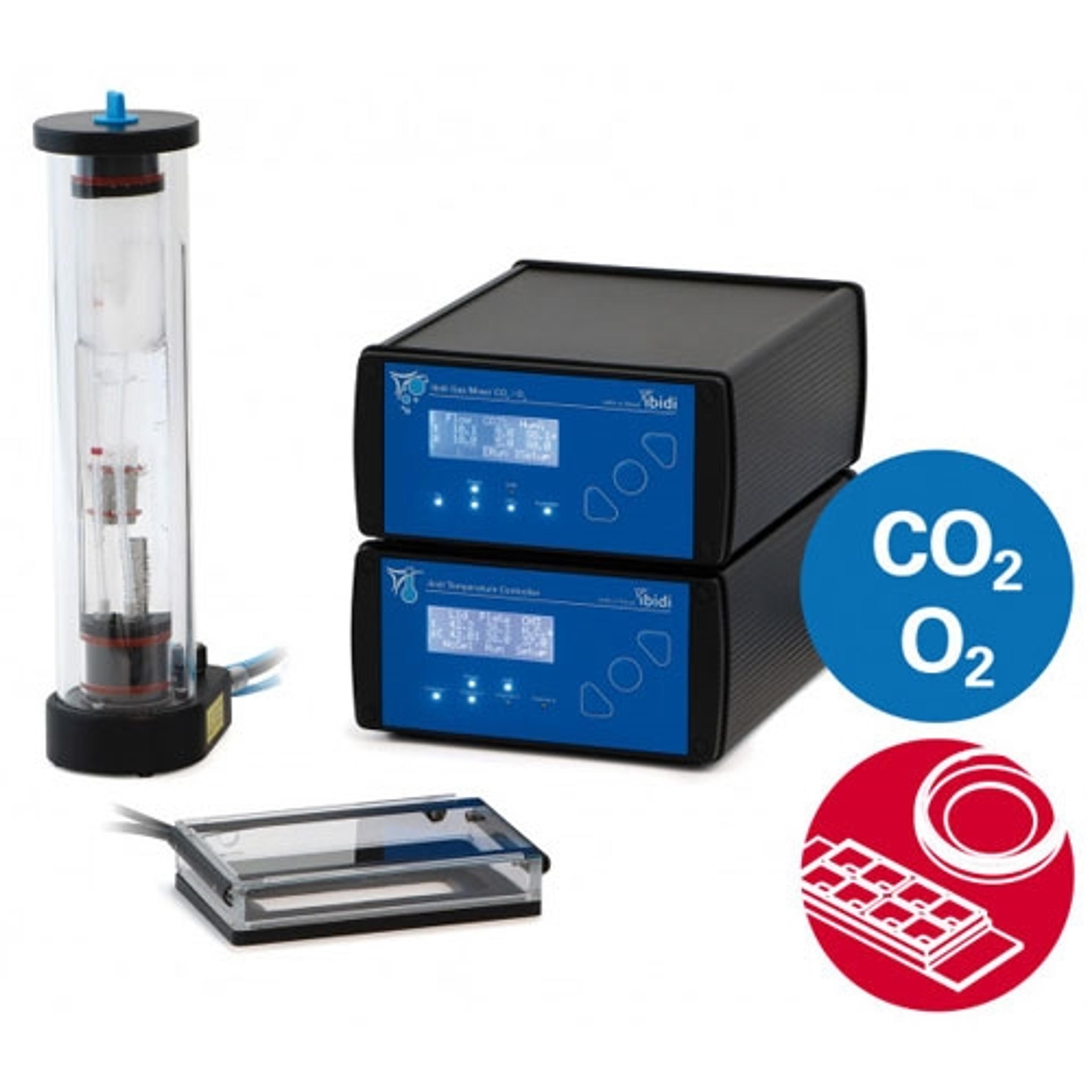
ibidi Stage Top Incubation System, with CO2 and O2 Control, for Live Cell Imaging
ibidi GmbHEasy setup directly on your microscope: perform live cell imaging in dishes, slides, or multiwell plates. The ibidi Stage Top Incubation Systems fit every standard inverted microscope and include CO 2 and O 2 control as well as actively controlled humidity. They are ideal for all live cell imaging applications and available for single slides and dishes as well as for multiwell plates.
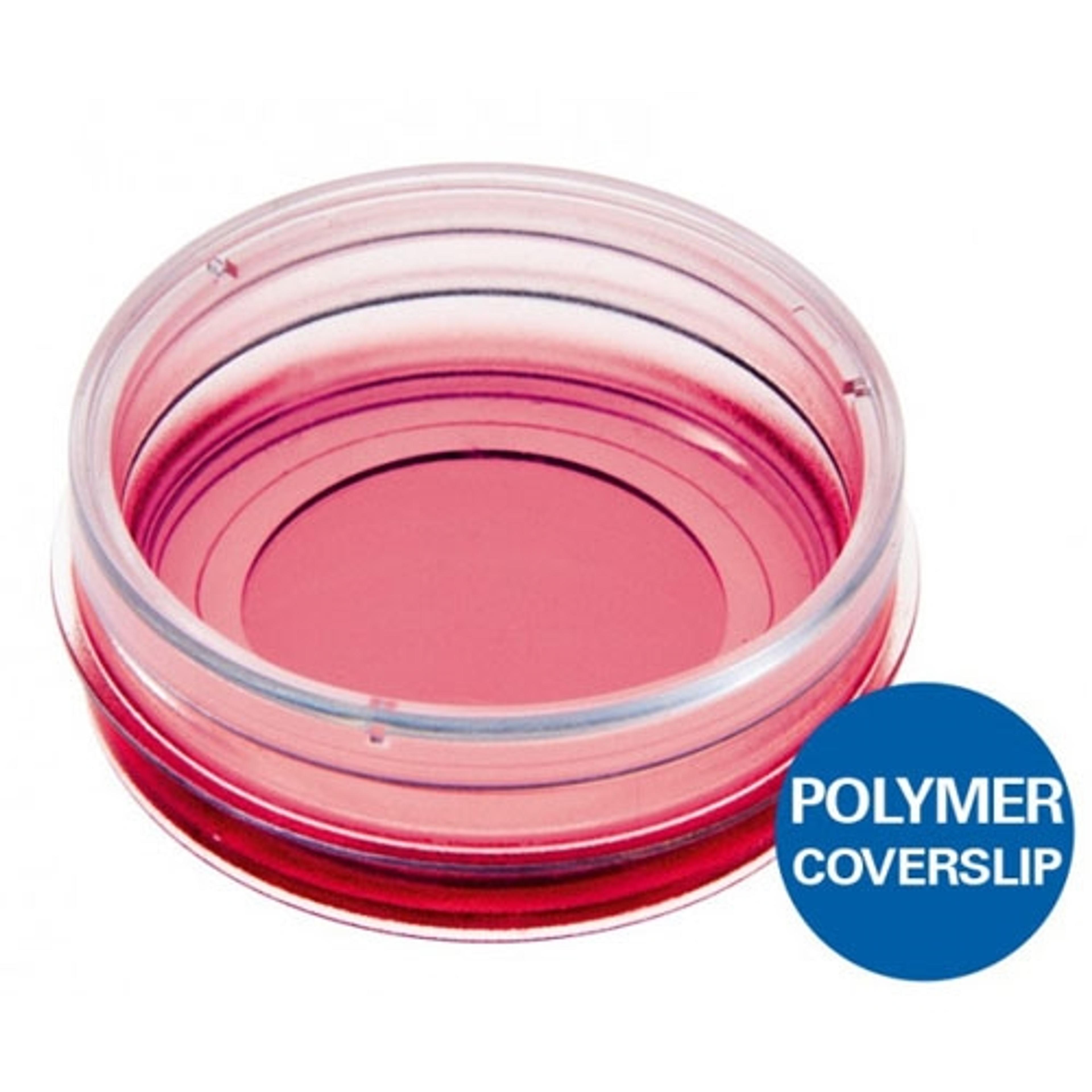
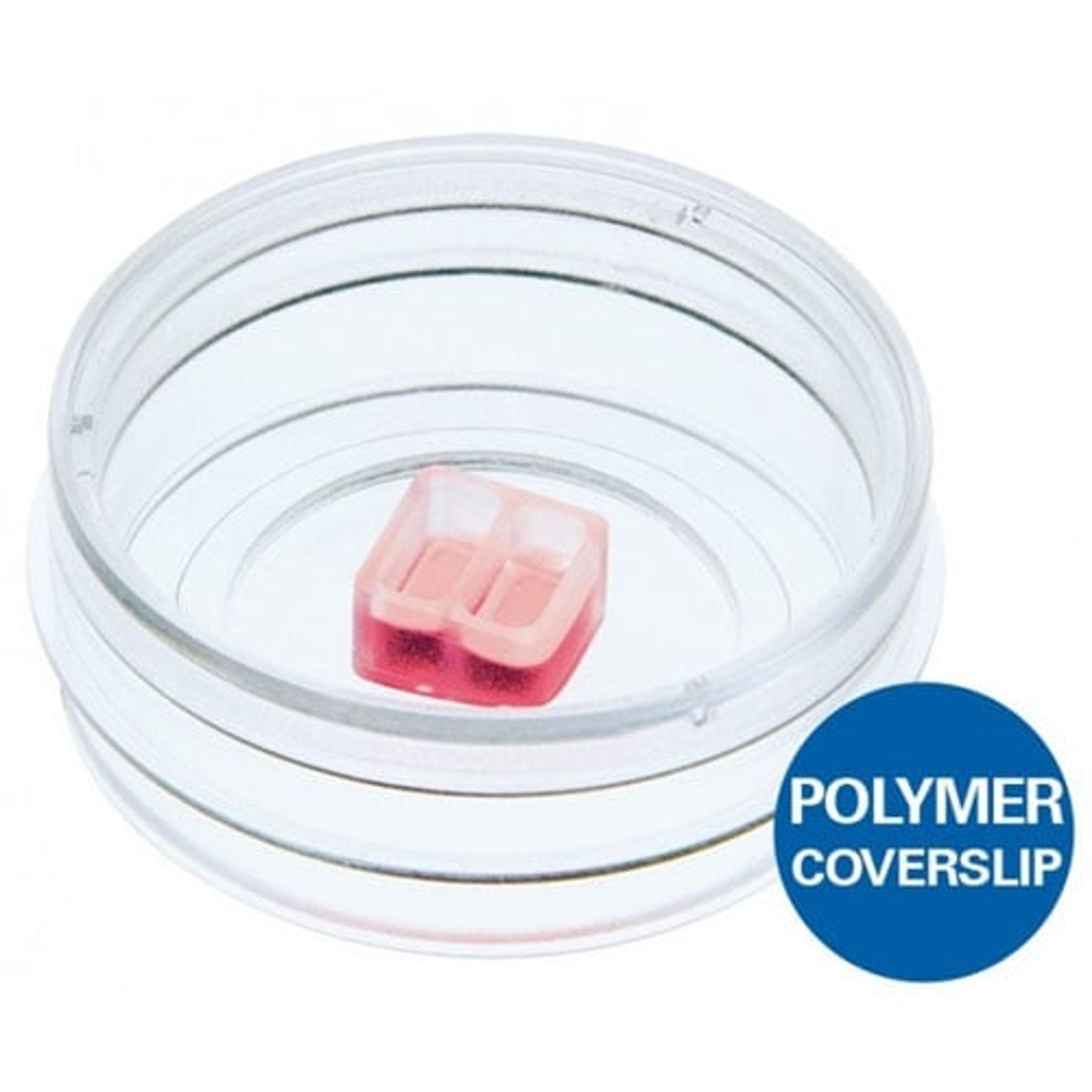
µ-Dish 35 mm, high
ibidi GmbHUse this versatile 35 mm imaging Dish for high-end microscopy through the #1.5 coverslip bottom.
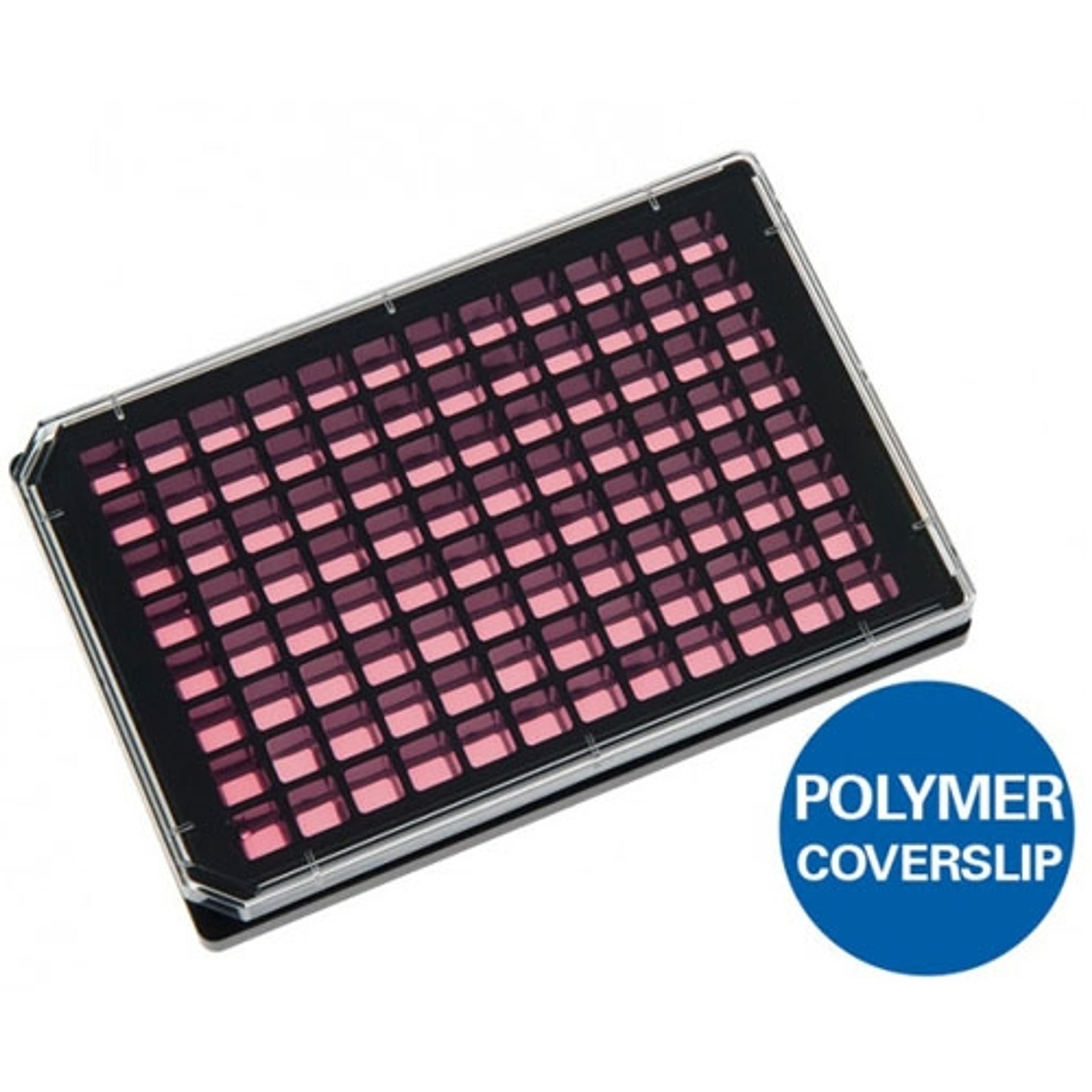
µ-Plate 96 Well Black
ibidi GmbHUse this Multiwell plate with black walls, square wells, and a flat and clear coverslip bottom (#1.5 ibidi Polymer Coverslip) for high-throughput applications in cell-based assays. Also available with 24 wells.
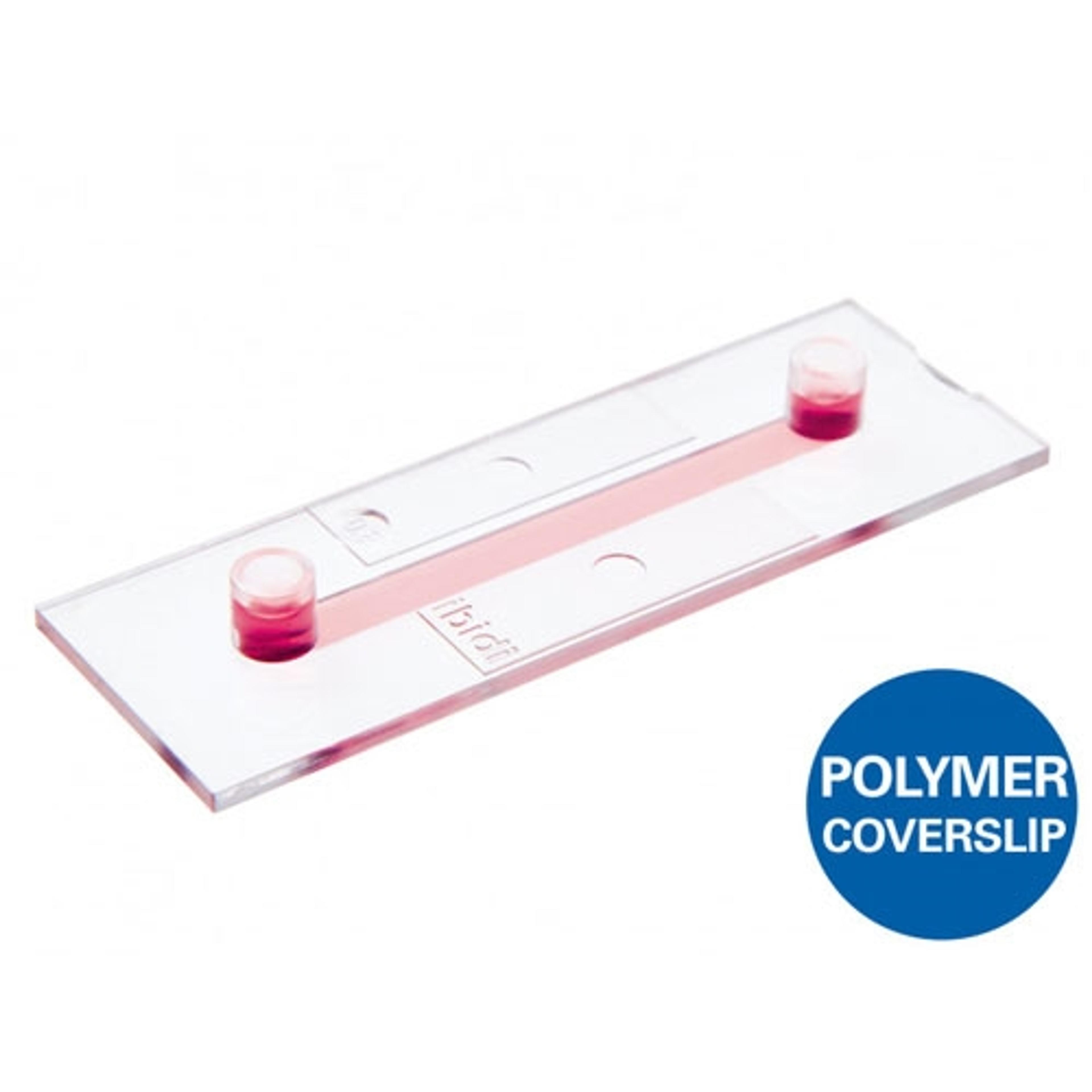
µ-Slide I Luer
ibidi GmbHUse this channel microscopy slide with a coverslip bottom (#1.5 ibidi Polymer or #1.5H glass) for immunofluorescence, cell culture under flow, live cell imaging, and high resolution microscopy on inverted microscopes. Available in various channel heights.
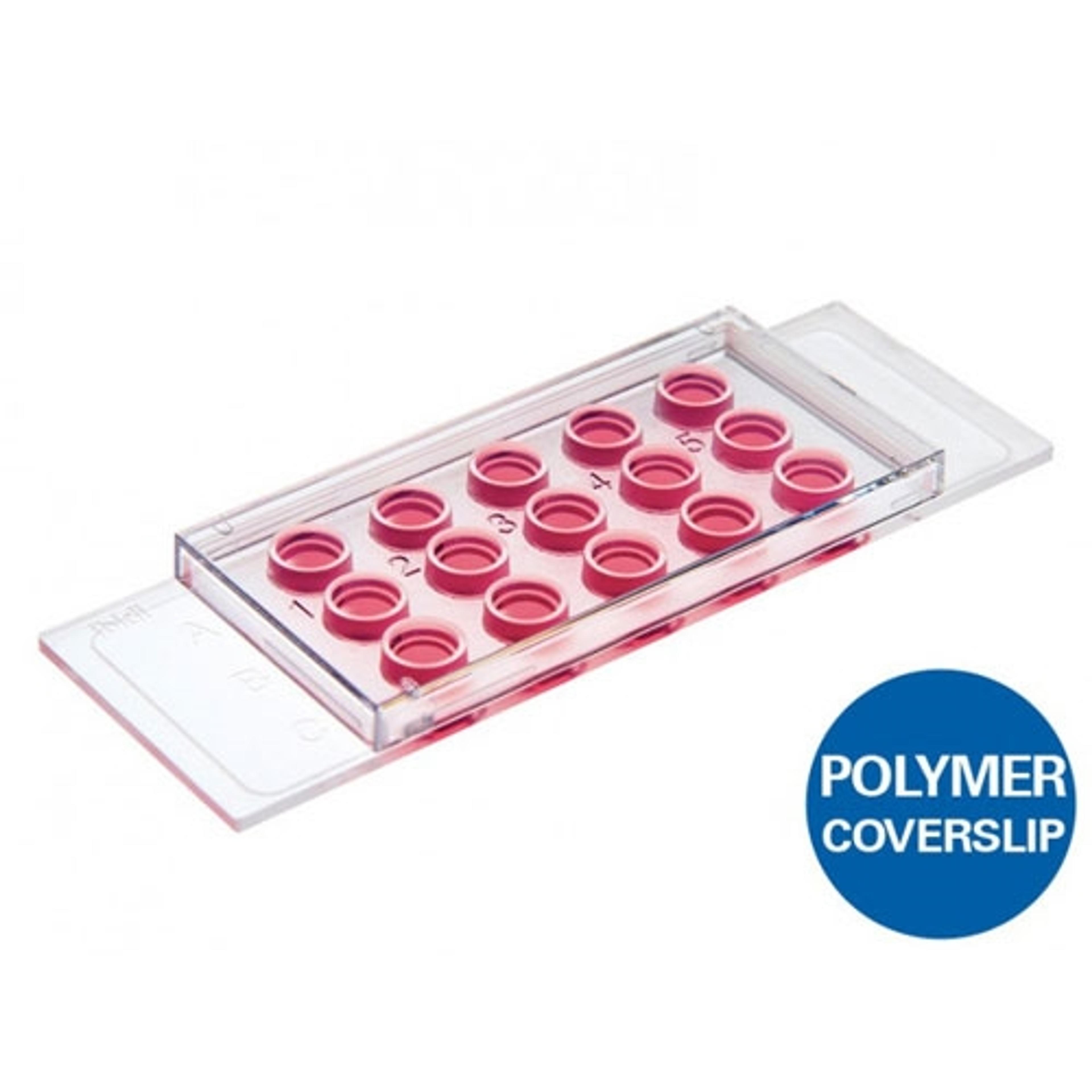
Culture-Insert 2 Well | 3 Well | 4 Well
ibidi GmbHUse these silicone inserts with a defined cell-free gap for wound healing assays, migration assays, 2D invasion assays, and co-cultivation of cells. Available with 2, 3, or 4 wells.
µ-Slide Angiogenesis
ibidi GmbHUse this slide to investigate angiogenesis in tube formation assays. Also ideal for 3D cell culture and immunofluorescence staining. Also available in a 96 well format.